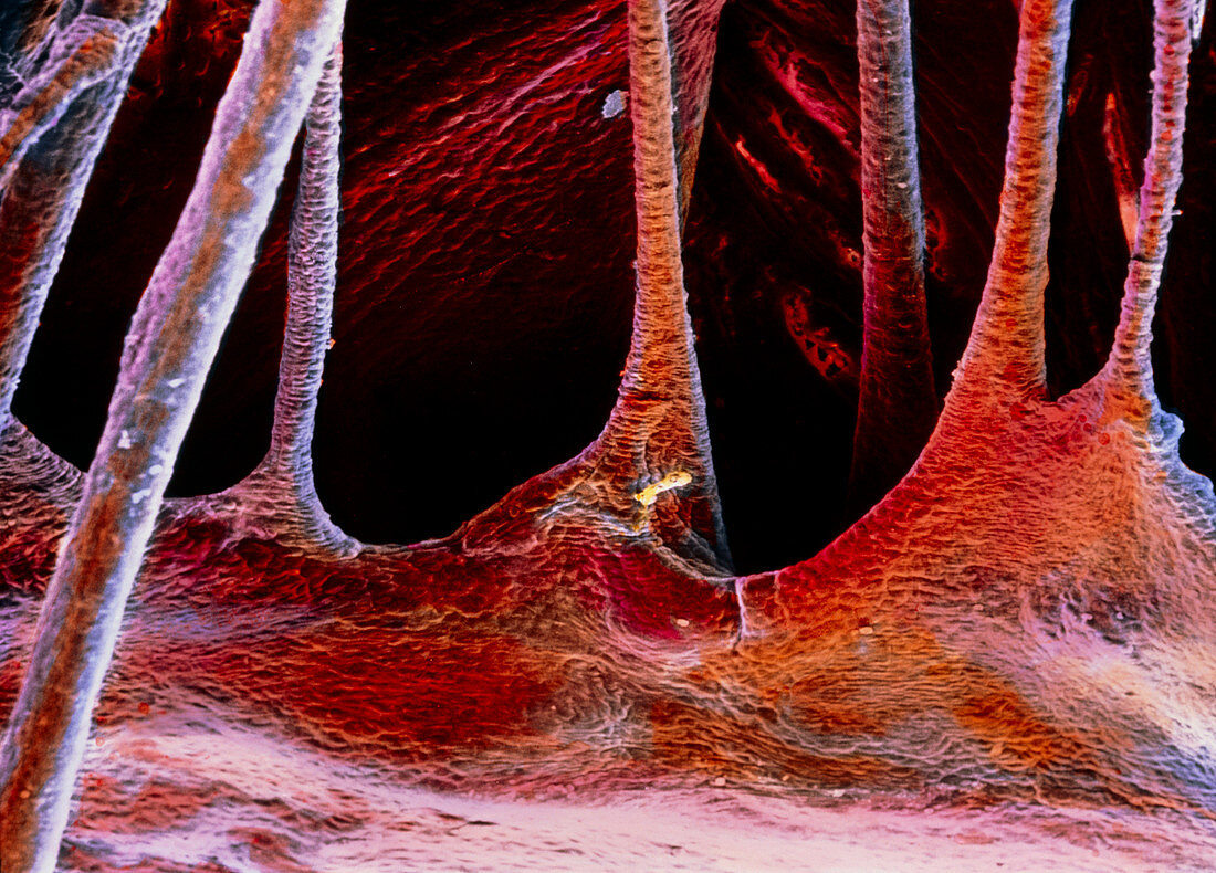
False-colour SEM of a portion of a cardiac valve
Bildnummer 11868987

| Heart valve. False-colour scanning electron micrograph of a portion of one of the atrioventricular valves of the heart: either the tricuspid or the mitral valve. They channel the flow of blood from the atria to the ventricles and prevent any backflow. The image clearly shows the chordae tendineae,the fibrous filiform collagenous cords,which connect the cusps of the valve to the papillary muscles of the heart. The epithelium at top is known as the endocardium: it is formed by a single layer of flattened cells supported by fibro-elastic connective tissue. Magnification: x70 at 6x7cm size. x115 at 4x5ins | |
| Lizenzart: | Lizenzpflichtig |
| Credit: | Science Photo Library / UNIVERSITY LA SAPIENZA, ROME / DEPT. OF ANATOMY / PROF. P. MOTTA |
| Bildgröße: | 5197 px × 3734 px |
| Modell-Rechte: | nicht erforderlich |
| Eigentums-Rechte: | nicht erforderlich |
| Restrictions: | - |
Preise für dieses Bild ab 15 €
Universitäten & Organisationen
(Informationsmaterial Digital, Informationsmaterial Print, Lehrmaterial Digital etc.)
ab 15 €
Redaktionell
(Bücher, Bücher: Sach- und Fachliteratur, Digitale Medien (redaktionell) etc.)
ab 30 €
Werbung
(Anzeigen, Aussenwerbung, Digitale Medien, Fernsehwerbung, Karten, Werbemittel, Zeitschriften etc.)
ab 55 €
Handelsprodukte
(bedruckte Textilie, Kalender, Postkarte, Grußkarte, Verpackung etc.)
ab 75 €
Pauschalpreise
Rechtepakete für die unbeschränkte Bildnutzung in Print oder Online
ab 495 €
