Haemangioblastoma brain tumour,MRA scan
Bildnummer 11839697
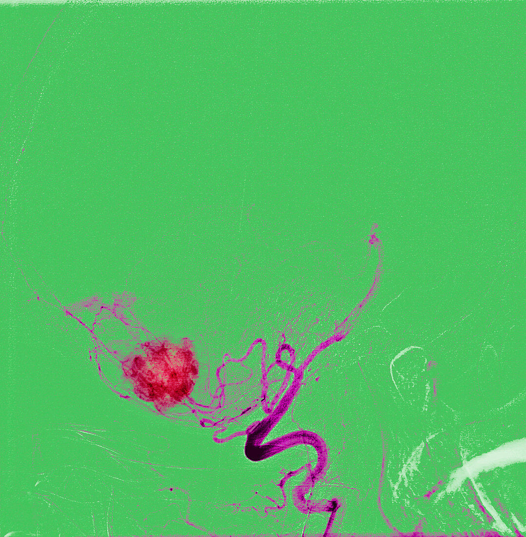
| Haemangioblastoma brain tumour. Coloured magnetic resonance angiography (MRA) scan of the brain of a 78 year old woman. The image shows the arterial blood supply (purple) to the haemangioblastoma tumour (red),which is affecting the left lobe of the cerebellum. Haemangoblastomas are formed from cells that line the blood vessels and are almost always benign. The main symptoms,such as headaches and sight problems,arise from increased pressure within the skull. Surgery is often the main form of treatment. MRA is a non-invasive technique to image blood vessels in the body that uses a combination of a very strong magnetic field and radiofrequency pulses to image the flow of blood | |
| Lizenzart: | Lizenzpflichtig |
| Credit: | Science Photo Library / Fraser, Simon |
| Bildgröße: | 2941 px × 2997 px |
| Modell-Rechte: | nicht erforderlich |
| Eigentums-Rechte: | nicht erforderlich |
| Restrictions: | - |
Preise für dieses Bild ab 15 €
Universitäten & Organisationen
(Informationsmaterial Digital, Informationsmaterial Print, Lehrmaterial Digital etc.)
ab 15 €
Redaktionell
(Bücher, Bücher: Sach- und Fachliteratur, Digitale Medien (redaktionell) etc.)
ab 30 €
Werbung
(Anzeigen, Aussenwerbung, Digitale Medien, Fernsehwerbung, Karten, Werbemittel, Zeitschriften etc.)
ab 55 €
Handelsprodukte
(bedruckte Textilie, Kalender, Postkarte, Grußkarte, Verpackung etc.)
ab 75 €
Pauschalpreise
Rechtepakete für die unbeschränkte Bildnutzung in Print oder Online
ab 495 €
Keywords
- Alt,
- älter,
- Angiografie,
- Angiogramm,
- Arterie,
- arteriell,
- Arterien,
- Blutgefäß,
- Blutgefäße,
- Diagnose,
- Erwachsene,
- farbig,
- Frau,
- gefärbt,
- Gehirn,
- gutartig,
- Hämangioblastom,
- Hirnscan,
- Kleinhirn,
- Kondition,
- Krankheit,
- Kreislauf,
- Magnetresonanzangiographie,
- Magnetresonanztomografie,
- Medizin,
- medizinisch,
- menschlicher Körper,
- MRA,
- MRT-Untersuchung,
- Neurologie,
- neurologisch,
- Onkologie,
- Scanner,
- siebziger Jahre,
- solide,
- Störung,
- Teilweise,
- Tumor,
- vaskulär,
- Wachstum,
- Weiblich,
- Zellen