Nerve,light micrograph
Bildnummer 11731389
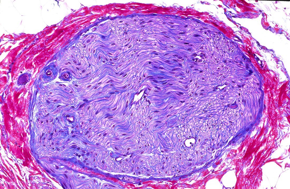
| Light microscopy of a small peripheral nerve in cross section. The hundreds of individual nerve fibres are seen in transverse,longitudinal and oblique profiles. Enclosed and supported by connective tissue (blue) the axons are generally unstained and many scattered nuclei (magenta) represent Schwann cells or fibroblasts. Peripheral blue-stained connective tissue margin is the perineurium providing support and protection. Magnification x150 when printed at 10 cm | |
| Lizenzart: | Lizenzpflichtig |
| Credit: | Science Photo Library / Microscape |
| Bildgröße: | 5172 px × 3378 px |
| Modell-Rechte: | nicht erforderlich |
| Eigentums-Rechte: | nicht erforderlich |
| Restrictions: | - |
Preise für dieses Bild ab 15 €
Universitäten & Organisationen
(Informationsmaterial Digital, Informationsmaterial Print, Lehrmaterial Digital etc.)
ab 15 €
Redaktionell
(Bücher, Bücher: Sach- und Fachliteratur, Digitale Medien (redaktionell) etc.)
ab 30 €
Werbung
(Anzeigen, Aussenwerbung, Digitale Medien, Fernsehwerbung, Karten, Werbemittel, Zeitschriften etc.)
ab 55 €
Handelsprodukte
(bedruckte Textilie, Kalender, Postkarte, Grußkarte, Verpackung etc.)
ab 75 €
Pauschalpreise
Rechtepakete für die unbeschränkte Bildnutzung in Print oder Online
ab 495 €