Child skeleton,18th century
Bildnummer 11727424
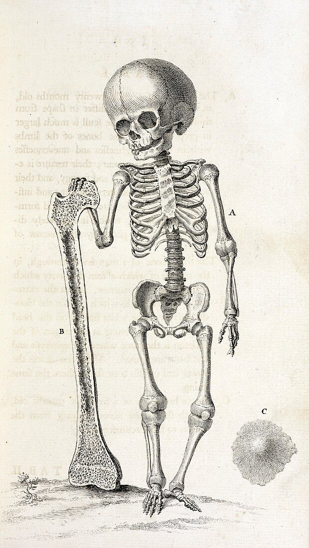
| Child skeleton. 18th-century illustration of the skeleton of a 20-month old baby holding an adult human male femur dissected in longitudinal section to shown the spongy and honeycombed interior. A foetal skull bone is shown at lower right. This artwork is from the fifth edition (1740) of 'The Anatomy of the Human Body' by English surgeon and anatomist William Cheselden (1688-1752). This book was first published in 1713 and was re-issued through 13 editions. It was popular due to being written in English instead of Latin | |
| Lizenzart: | Lizenzpflichtig |
| Credit: | Science Photo Library / British Library |
| Bildgröße: | 3152 px × 5596 px |
| Modell-Rechte: | nicht erforderlich |
| Eigentums-Rechte: | nicht erforderlich |
| Restrictions: | - |
Preise für dieses Bild ab 15 €
Universitäten & Organisationen
(Informationsmaterial Digital, Informationsmaterial Print, Lehrmaterial Digital etc.)
ab 15 €
Redaktionell
(Bücher, Bücher: Sach- und Fachliteratur, Digitale Medien (redaktionell) etc.)
ab 30 €
Werbung
(Anzeigen, Aussenwerbung, Digitale Medien, Fernsehwerbung, Karten, Werbemittel, Zeitschriften etc.)
ab 55 €
Handelsprodukte
(bedruckte Textilie, Kalender, Postkarte, Grußkarte, Verpackung etc.)
ab 75 €
Pauschalpreise
Rechtepakete für die unbeschränkte Bildnutzung in Print oder Online
ab 495 €
Keywords
- 1700er Jahre,
- 18. Jahrhundert,
- Anatomie,
- anatomisch,
- Baby,
- Bein,
- Blatt,
- Buch,
- Einfarbig,
- Femur,
- fötal,
- Fötus,
- ganzer Körper,
- Geschichte,
- gesund,
- historisch,
- Illustration,
- Innere,
- innere Struktur,
- Kind,
- Knochen,
- Knochenmark,
- Kunstwerk,
- menschlicher Körper,
- Niemand,
- normal,
- Oberschenkelknochen,
- Osteologie,
- Säugling,
- Schädel,
- Schwarz und weiß,
- Sektion,
- sektioniert,
- Skelett,
- Veröffentlichung,
- William Chesleden