Femur bones,18th century
Bildnummer 11725795
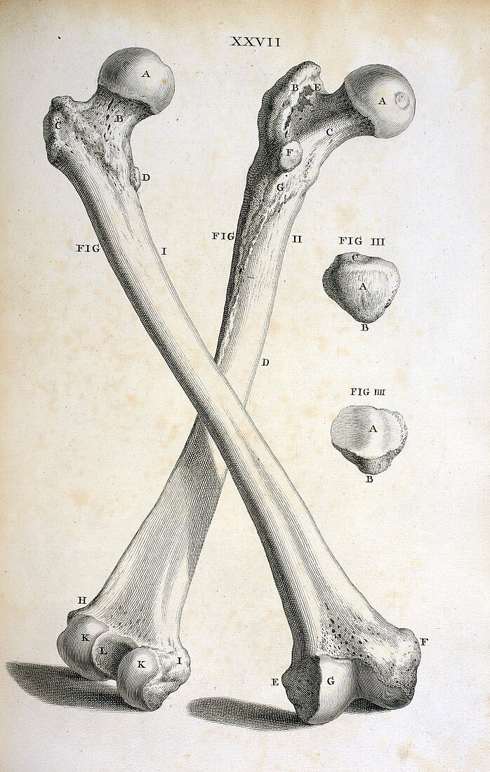
| Femur bones. 18th-century illustration of human femurs (thigh bones) and patellae (kneecaps). The right femur is shown at the front and the left behind. The balls at the top of the femurs fit into the hip sockets. At the bottom are the upper halves of the hinge joints that form the knees. The patellae (centre right) protect the front of the knee joints. This illustration is from 'Osteographia,or the Anatomy of the bones' (London,1733) by English surgeon and anatomist William Cheselden (1688-1752). It was the first full and accurate description of the anatomy of the human skeletal system | |
| Lizenzart: | Lizenzpflichtig |
| Credit: | Science Photo Library / British Library |
| Bildgröße: | 3337 px × 5258 px |
| Modell-Rechte: | nicht erforderlich |
| Eigentums-Rechte: | nicht erforderlich |
| Restrictions: | - |
Preise für dieses Bild ab 15 €
Universitäten & Organisationen
(Informationsmaterial Digital, Informationsmaterial Print, Lehrmaterial Digital etc.)
ab 15 €
Redaktionell
(Bücher, Bücher: Sach- und Fachliteratur, Digitale Medien (redaktionell) etc.)
ab 30 €
Werbung
(Anzeigen, Aussenwerbung, Digitale Medien, Fernsehwerbung, Karten, Werbemittel, Zeitschriften etc.)
ab 55 €
Handelsprodukte
(bedruckte Textilie, Kalender, Postkarte, Grußkarte, Verpackung etc.)
ab 75 €
Pauschalpreise
Rechtepakete für die unbeschränkte Bildnutzung in Print oder Online
ab 495 €
Keywords
- 1700er Jahre,
- 18. Jahrhundert,
- Anatomie,
- Anatomie der Knochen,
- anatomisch,
- Blatt,
- Buch,
- Einfarbig,
- England,
- Englisch,
- europäisch,
- Femur,
- Geschichte,
- gesund,
- historisch,
- Illustration,
- Kappen,
- Knochen,
- Kunstwerk,
- links,
- Medizin,
- medizinisch,
- Niemand,
- normal,
- Oberschenkelknochen,
- Osteologie,
- Recht,
- Schwarz und weiß,
- Skelett-,
- Veröffentlichung,
- William Cheselden