Bone and cartilage,light micrograph
Bildnummer 11704753
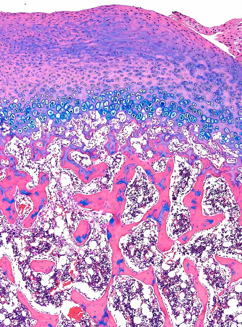
| Light microscopy of developing bone. The top layer (purple,pink) is cartilage which covers the end of the bone; next layer (blue) is the epiphyseal growth plate comprising chondrocytes; lower half shows spongy bone (pink) of honeycomb appearance with remnants of calcifying cartilage (blue) surrounded by bone tissue. The gaps between the spongy bone is occupied by bone marrow cells. Magnification x90 when narrow width printed at 10 cm | |
| Lizenzart: | Lizenzpflichtig |
| Credit: | Science Photo Library / Microscape |
| Bildgröße: | 3629 px × 4890 px |
| Modell-Rechte: | nicht erforderlich |
| Eigentums-Rechte: | nicht erforderlich |
| Restrictions: | - |
Preise für dieses Bild ab 15 €
Universitäten & Organisationen
(Informationsmaterial Digital, Informationsmaterial Print, Lehrmaterial Digital etc.)
ab 15 €
Redaktionell
(Bücher, Bücher: Sach- und Fachliteratur, Digitale Medien (redaktionell) etc.)
ab 30 €
Werbung
(Anzeigen, Aussenwerbung, Digitale Medien, Fernsehwerbung, Karten, Werbemittel, Zeitschriften etc.)
ab 55 €
Handelsprodukte
(bedruckte Textilie, Kalender, Postkarte, Grußkarte, Verpackung etc.)
ab 75 €
Pauschalpreise
Rechtepakete für die unbeschränkte Bildnutzung in Print oder Online
ab 495 €