Brain in Creutzfeldt-Jakob disease,MRI
Bildnummer 11704620
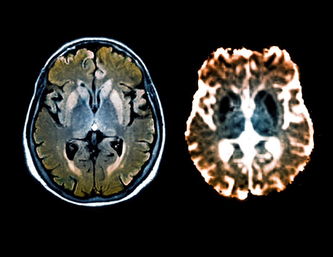
| Brain in Creutzfeldt-Jakob disease. FLAIR and diffusion magnetic resonance imaging (MRI) scans of a section through the brain of a 47-old patient,showing hyperintensities around the area of the cortex and basal ganglia,suggestive of Creutzfeldt-Jakob disease (CJD). CJD is the result of virus-like prions (misfolded proteins) within the brain that cause vacuoles and plaques to form,making the brain spongy and killing off the tissue. Symptoms include dementia and sudden muscle contractions,leading to death. The similarly fatal cattle disease BSE (bovine spongiform encephalopathy) has now been linked to human CJD | |
| Lizenzart: | Lizenzpflichtig |
| Credit: | Science Photo Library / Zephyr |
| Bildgröße: | 4014 px × 3103 px |
| Modell-Rechte: | nicht erforderlich |
| Eigentums-Rechte: | nicht erforderlich |
| Restrictions: | - |
Preise für dieses Bild ab 15 €
Universitäten & Organisationen
(Informationsmaterial Digital, Informationsmaterial Print, Lehrmaterial Digital etc.)
ab 15 €
Redaktionell
(Bücher, Bücher: Sach- und Fachliteratur, Digitale Medien (redaktionell) etc.)
ab 30 €
Werbung
(Anzeigen, Aussenwerbung, Digitale Medien, Fernsehwerbung, Karten, Werbemittel, Zeitschriften etc.)
ab 55 €
Handelsprodukte
(bedruckte Textilie, Kalender, Postkarte, Grußkarte, Verpackung etc.)
ab 75 €
Pauschalpreise
Rechtepakete für die unbeschränkte Bildnutzung in Print oder Online
ab 495 €
Keywords
- abnormal,
- Abschnitte,
- Anatomie,
- anatomisch,
- ausgeschnitten,
- Ausschnitte,
- Basalganglien,
- Cortex,
- Creutzfeldt-Jakob,
- Creutzfeldt-Jakob-Krankheit,
- diagnostische Bildgebung,
- farbig,
- Flair,
- gefärbt,
- Gehirn,
- Gesundheitswesen,
- Hyperintensität,
- Kondition,
- Kopf,
- krank,
- Krankheit,
- Magnetresonanztomografie,
- Medizin,
- medizinisch,
- menschlicher Körper,
- MRI,
- Neuroimaging,
- Neurologie,
- neurologisch,
- Niemand,
- Organ,
- Plague,
- Prion,
- Radiographie,
- Radiologie,
- radiologisch,
- Scan,
- schwarzer Hintergrund,
- Sektion,
- sektioniert,
- Störung,
- ungesund,
- zentrales Nervensystem