32 week foetus,3-D ultrasound scan
Bildnummer 11645035
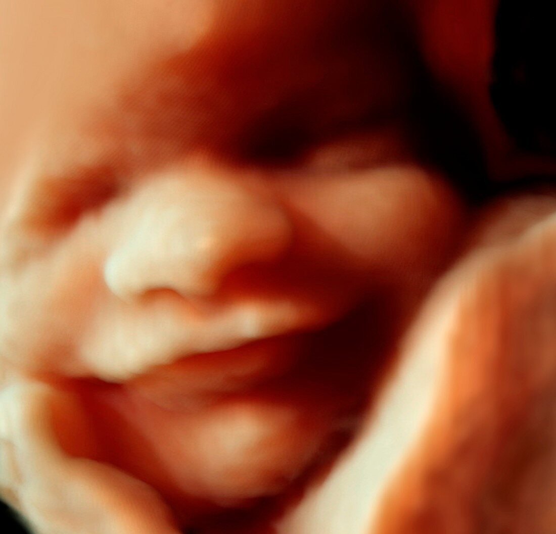
| 32 week foetus,3-D ultrasound scan. Three-dimensional (3-D) ultrasound scan of a human foetus. The image was produced by a 3-D ultrasound scanner called Voluson E8 utilising software called HDLive. 3-D scanning enables physiological disorders such as harelip and spina bifida to be diagnosed before birth. Ultrasound is a diagnostic technique which sends high-frequency sound waves into the body via a transducer. The returning echoes are recorded and used to build an image of an internal structure. HDLive renders the ultrasound data in real time with realistic lighting and surface detail | |
| Lizenzart: | Lizenzpflichtig |
| Credit: | Science Photo Library / Benoit, Bernard |
| Bildgröße: | 3012 px × 2895 px |
| Modell-Rechte: | Derzeit liegt noch kein Release vor. Bitte kontaktieren Sie uns vor Verwendung. |
| Eigentums-Rechte: | nicht erforderlich |
| Restrictions: | - |
Preise für dieses Bild ab 15 €
Universitäten & Organisationen
(Informationsmaterial Digital, Informationsmaterial Print, Lehrmaterial Digital etc.)
ab 15 €
Redaktionell
(Bücher, Bücher: Sach- und Fachliteratur, Digitale Medien (redaktionell) etc.)
ab 30 €
Werbung
(Anzeigen, Aussenwerbung, Digitale Medien, Fernsehwerbung, Karten, Werbemittel, Zeitschriften etc.)
ab 55 €
Handelsprodukte
(bedruckte Textilie, Kalender, Postkarte, Grußkarte, Verpackung etc.)
ab 75 €
Pauschalpreise
Rechtepakete für die unbeschränkte Bildnutzung in Print oder Online
ab 495 €
Keywords
- 2013,
- 3-d,
- 3D,
- Auge,
- Augenlid,
- Baby,
- Baby-Scan,
- Bild,
- Biologie,
- biologisch,
- Diagnose,
- Dreidimensional,
- Entwicklung,
- Fötus,
- Gebärmutter,
- Geburtshilfe,
- Gesicht,
- Gesundheitswesen,
- hdlive,
- Kopf,
- Lächeln,
- Lächelnd,
- Lippe,
- Lippen,
- Medizin,
- medizinisch,
- Mensch,
- Menschen,
- menschlicher Körper,
- Mund,
- Nase,
- Pädiatrie,
- Person,
- Reproduktion,
- Säugling,
- Scan,
- Scanner,
- Schallwellen,
- Schwangerschaft,
- Sonographie,
- Ultraschall,
- Ultraschalluntersuchung,
- ungeboren,
- Voluson,
- Voluson E8