F/col STM image of HPOG graphite surface
Bildnummer 11518373
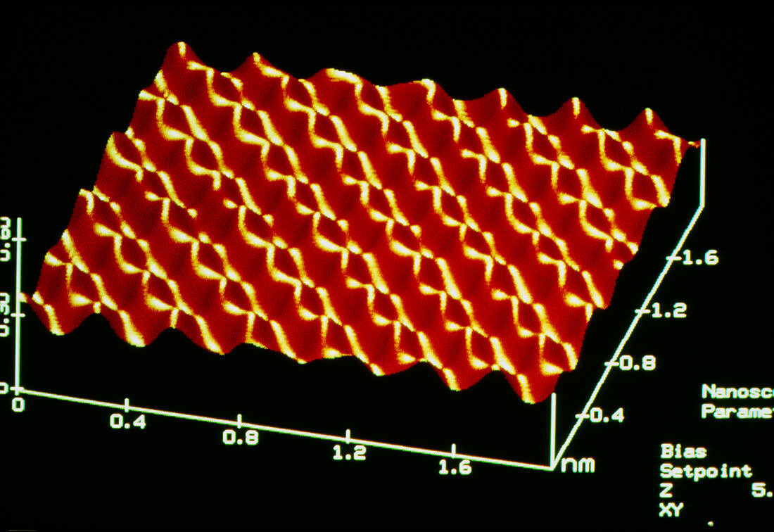
| False-colour scanning tunnelling microscope (STM) image of the surface of a sample of Highly- Ordered Pyrolitic Graphite (HPOG),revealing the regular pattern of individual carbon atoms. The STM provides a detailed image of the surfaces of certain samples,allowing superficial atoms to be identified. Basically,the image is formed by moving a fine point just above the sample surface & electronically recording the height of the point as it scans. This is possible due to tunnelling,whereby an exchange of electrons occurs when the electron clouds surrounding superficial nuclei in the STM's point & sample surface overlap as they approach each other | |
| Lizenzart: | Lizenzpflichtig |
| Credit: | Science Photo Library / Plailly, Philippe |
| Bildgröße: | 5428 px × 3730 px |
| Modell-Rechte: | nicht erforderlich |
| Eigentums-Rechte: | nicht erforderlich |
| Restrictions: |
|
Preise für dieses Bild ab 15 €
Universitäten & Organisationen
(Informationsmaterial Digital, Informationsmaterial Print, Lehrmaterial Digital etc.)
ab 15 €
Redaktionell
(Bücher, Bücher: Sach- und Fachliteratur, Digitale Medien (redaktionell) etc.)
ab 30 €
Werbung
(Anzeigen, Aussenwerbung, Digitale Medien, Fernsehwerbung, Karten, Werbemittel, Zeitschriften etc.)
ab 55 €
Handelsprodukte
(bedruckte Textilie, Kalender, Postkarte, Grußkarte, Verpackung etc.)
ab 75 €
Pauschalpreise
Rechtepakete für die unbeschränkte Bildnutzung in Print oder Online
ab 495 €