Large bowel, light micrograph
Bildnummer 13633597
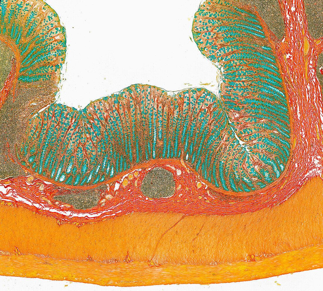
| Light micrograph of the large bowel (colon) showing the mucosa (green), submucosa (orange) and muscularis externa (yellow). The mucosa has elongated tubular glands with mucus-secreting goblet cells stained green. Loose connective tissue of the submucosa shows occasional lymphoid aggregations (grey stain). The muscularis externa consists of two layers of smooth muscle an inner circular and an outer longitudinal layer. Paraffin section, alcian blue and van Gieson's stain. Magnification: x40 when width printed at 10cm. | |
| Lizenzart: | Lizenzpflichtig |
| Credit: | Science Photo Library / Microscape |
| Bildgröße: | 4724 px × 4256 px |
| Modell-Rechte: | nicht erforderlich |
| Eigentums-Rechte: | nicht erforderlich |
| Restrictions: | - |
Preise für dieses Bild ab 15 €
Universitäten & Organisationen
(Informationsmaterial Digital, Informationsmaterial Print, Lehrmaterial Digital etc.)
ab 15 €
Redaktionell
(Bücher, Bücher: Sach- und Fachliteratur, Digitale Medien (redaktionell) etc.)
ab 30 €
Werbung
(Anzeigen, Aussenwerbung, Digitale Medien, Fernsehwerbung, Karten, Werbemittel, Zeitschriften etc.)
ab 55 €
Handelsprodukte
(bedruckte Textilie, Kalender, Postkarte, Grußkarte, Verpackung etc.)
ab 75 €
Pauschalpreise
Rechtepakete für die unbeschränkte Bildnutzung in Print oder Online
ab 495 €