Arteriole, light micrograph
Bildnummer 13633589
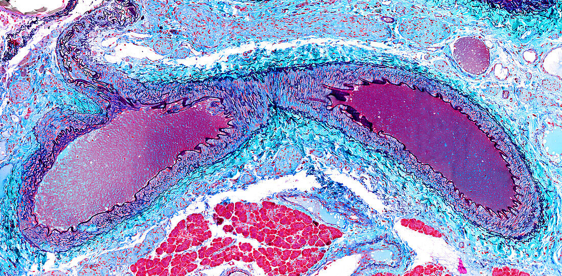
| Light micrograph of a longitudinal section of an arteriole. The wall (tunica media) is composed of smooth muscle cells with elastic fibres (purple-black stain). Facing the lumen filled with blood cells and plasma (stained magenta) is the folded internal elastic lamina. External to the tunica media is the tunica adventitia of connective tissue stained green. The external elastic lamina lies between the two tunics. A small branching process is at upper left. Paraffin section, Gomori trichrome stain. Magnification: x75 when width printed at 10cm. | |
| Lizenzart: | Lizenzpflichtig |
| Credit: | Science Photo Library / Microscape |
| Bildgröße: | 6688 px × 3288 px |
| Modell-Rechte: | nicht erforderlich |
| Eigentums-Rechte: | nicht erforderlich |
| Restrictions: | - |
Preise für dieses Bild ab 15 €
Universitäten & Organisationen
(Informationsmaterial Digital, Informationsmaterial Print, Lehrmaterial Digital etc.)
ab 15 €
Redaktionell
(Bücher, Bücher: Sach- und Fachliteratur, Digitale Medien (redaktionell) etc.)
ab 30 €
Werbung
(Anzeigen, Aussenwerbung, Digitale Medien, Fernsehwerbung, Karten, Werbemittel, Zeitschriften etc.)
ab 55 €
Handelsprodukte
(bedruckte Textilie, Kalender, Postkarte, Grußkarte, Verpackung etc.)
ab 75 €
Pauschalpreise
Rechtepakete für die unbeschränkte Bildnutzung in Print oder Online
ab 495 €