Spermatogenesis in human testicle, light micrographs
Bildnummer 13613990
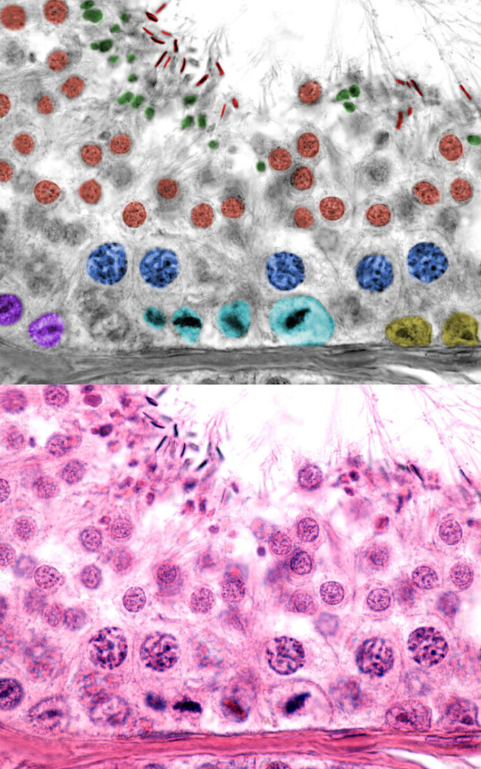
| Human testicle, light micrographs. The bottom micrograph shows a seminiferous tubule. In the top micrograph the cell types of the male germinal epithelium have been marked with colour. Seen are; Sertoli cells (yellow), spermatogonia (pink), spermatogonia in metaphase (light blue), primary spermatocytes in pachytene phase (blue), spermatids (brown), spermatozoa (red) and residual bodies (green). | |
| Lizenzart: | Lizenzpflichtig |
| Credit: | Science Photo Library / JOSE CALVO |
| Bildgröße: | 3840 px × 6144 px |
| Modell-Rechte: | nicht erforderlich |
| Eigentums-Rechte: | nicht erforderlich |
| Restrictions: | - |
Preise für dieses Bild ab 15 €
Universitäten & Organisationen
(Informationsmaterial Digital, Informationsmaterial Print, Lehrmaterial Digital etc.)
ab 15 €
Redaktionell
(Bücher, Bücher: Sach- und Fachliteratur, Digitale Medien (redaktionell) etc.)
ab 30 €
Werbung
(Anzeigen, Aussenwerbung, Digitale Medien, Fernsehwerbung, Karten, Werbemittel, Zeitschriften etc.)
ab 55 €
Handelsprodukte
(bedruckte Textilie, Kalender, Postkarte, Grußkarte, Verpackung etc.)
ab 75 €
Pauschalpreise
Rechtepakete für die unbeschränkte Bildnutzung in Print oder Online
ab 495 €