Vitreomacular traction, fundoscopy
Bildnummer 13613653
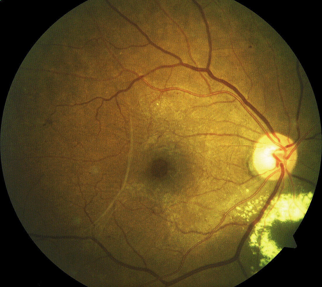
| Fundoscopy image of the retina of the right eye of a female patient with vitreomacular traction (VMT) and a retinal macroaneurysm. As part of the normal aging process the vitreous humour, the clear gel that fills the eyeball, shrinks and pulls away from the retina, the light-sensitive layer lining the inner surface of the eye. Eventually it will separate from the retina entirely. In cases of VMT part of the vitreous remains attached to the centre of the retina, known as the macula. This vitreous pulls on the macula, causing damage that can lead to distorted vision. Retinal macroaneurysm is the dilation of a retinal artery. Severe cases of VMT can be corrected with surgery. | |
| Lizenzart: | Lizenzpflichtig |
| Credit: | Science Photo Library / ALAN FROHLICHSTEIN |
| Bildgröße: | 3447 px × 3073 px |
| Modell-Rechte: | nicht erforderlich |
| Eigentums-Rechte: | nicht erforderlich |
| Restrictions: | - |
Preise für dieses Bild ab 15 €
Universitäten & Organisationen
(Informationsmaterial Digital, Informationsmaterial Print, Lehrmaterial Digital etc.)
ab 15 €
Redaktionell
(Bücher, Bücher: Sach- und Fachliteratur, Digitale Medien (redaktionell) etc.)
ab 30 €
Werbung
(Anzeigen, Aussenwerbung, Digitale Medien, Fernsehwerbung, Karten, Werbemittel, Zeitschriften etc.)
ab 55 €
Handelsprodukte
(bedruckte Textilie, Kalender, Postkarte, Grußkarte, Verpackung etc.)
ab 75 €
Pauschalpreise
Rechtepakete für die unbeschränkte Bildnutzung in Print oder Online
ab 495 €
Keywords
- abnormal,
- Altern,
- alternd,
- Arterie,
- arteriell,
- Auge,
- Augenheilkunde,
- Augenspiegel,
- Beschädigt,
- Diagnose,
- Farbe,
- Frau,
- Futter,
- Gesundheitswesen,
- Humor,
- Innere,
- Kondition,
- Makula,
- Medizin,
- medizinisch,
- menschlicher Körper,
- Netzhaut,
- Netzhaut-,
- Niemand,
- Oberfläche,
- ophthalmologisch,
- Ophthalmoskopie,
- optisch,
- Retina,
- Schaden,
- Sektion,
- sektioniert,
- Sicht,
- Störung,
- ungesund,
- Vision,
- Weiblich,
- Ziehen