Smoker's lungs with oat cell carcinoma, illustration
Bildnummer 13601102
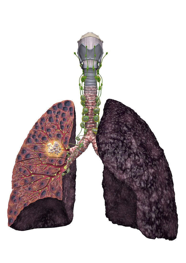
| Illustration of the lungs of a smoker with an oat cell carcinoma (white). The surface of the left lung (right) is darkened by chemicals from smoking cigarettes. The right lung (left) is shown in cross-section and reveals the inflamed red walls of the bronchi (tubes). The bronchi are lined with hair-like cilia, which beat to remove particles from the lungs. However, the toxic particles in cigarette smoke kill the cilia, leading to an increase in the number of mucus producing cells. This extra mucus now blocks the airways leading to inflammation and destruction of the alveoli (air sacs), which inflate and leave dark black holes. The destruction of the alveoli is irreversible, and decreases the lung function. Oat cell carcinoma is an aggressive cancer with a poor prognosis. | |
| Lizenzart: | Lizenzpflichtig |
| Credit: | Science Photo Library / Michael Alexowski |
| Bildgröße: | 3519 px × 4980 px |
| Modell-Rechte: | nicht erforderlich |
| Eigentums-Rechte: | nicht erforderlich |
| Restrictions: | - |
Preise für dieses Bild ab 15 €
Universitäten & Organisationen
(Informationsmaterial Digital, Informationsmaterial Print, Lehrmaterial Digital etc.)
ab 15 €
Redaktionell
(Bücher, Bücher: Sach- und Fachliteratur, Digitale Medien (redaktionell) etc.)
ab 30 €
Werbung
(Anzeigen, Aussenwerbung, Digitale Medien, Fernsehwerbung, Karten, Werbemittel, Zeitschriften etc.)
ab 55 €
Handelsprodukte
(bedruckte Textilie, Kalender, Postkarte, Grußkarte, Verpackung etc.)
ab 75 €
Pauschalpreise
Rechtepakete für die unbeschränkte Bildnutzung in Print oder Online
ab 495 €
Keywords
- 3D,
- Alveolen,
- Anatomie,
- anatomisch,
- Bronchien,
- Bronchus,
- cgi,
- chronisch obstruktive Lungenerkrankung,
- COPD,
- digital generiert,
- Entzündung,
- Illustration,
- kleinzelliges Karzinom,
- Krebs,
- Kunstwerk,
- Lunge,
- Lungen,
- Lungenkrebs,
- medizinisch,
- Metastase,
- Metastasen,
- Niemand,
- Onkologie,
- Rauchen,
- Raucher,
- Tumor,
- Zerstörung