Firearm injury, CT scan
Bildnummer 13599489
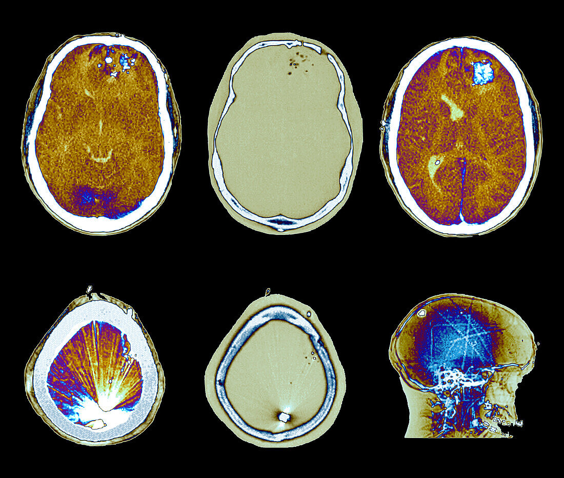
| Computed tomography (CT) and X-ray scans in axial sections (parenchymal and bone windows) of a 37-year-old patient admitted to the neurosurgical emergency room for firearm injury. The scan shows the presence of multiple bone fragments and projectiles in the left fronto-encephalic territory, with an entrance breach in the left frontal bone, presence of left hemispheric cerebral oedema, with mass effect on the ventricular structures, and arctefactant hyperdensity corresponding to the projectile impacted at the level posterior-high of the bony cranial box. | |
| Lizenzart: | Lizenzpflichtig |
| Credit: | Science Photo Library / Zephyr |
| Bildgröße: | 4017 px × 3402 px |
| Modell-Rechte: | nicht erforderlich |
| Eigentums-Rechte: | nicht erforderlich |
| Restrictions: | - |
Preise für dieses Bild ab 15 €
Universitäten & Organisationen
(Informationsmaterial Digital, Informationsmaterial Print, Lehrmaterial Digital etc.)
ab 15 €
Redaktionell
(Bücher, Bücher: Sach- und Fachliteratur, Digitale Medien (redaktionell) etc.)
ab 30 €
Werbung
(Anzeigen, Aussenwerbung, Digitale Medien, Fernsehwerbung, Karten, Werbemittel, Zeitschriften etc.)
ab 55 €
Handelsprodukte
(bedruckte Textilie, Kalender, Postkarte, Grußkarte, Verpackung etc.)
ab 75 €
Pauschalpreise
Rechtepakete für die unbeschränkte Bildnutzung in Print oder Online
ab 495 €