Peroxisomes in kidney convoluted tubule, light micrograph
Bildnummer 13586061
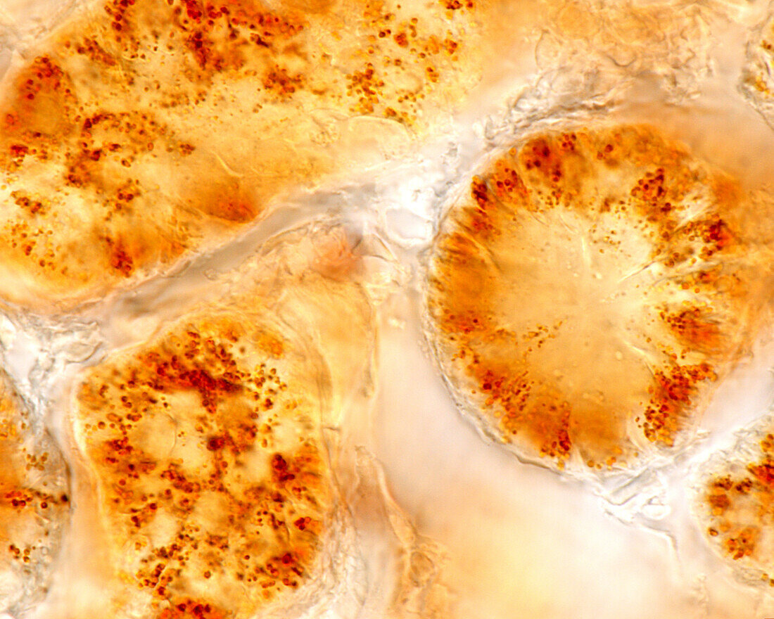
| Light micrograph of a kidney stained with the diaminobenzidine method to show peroxidase present in the peroxisomes of the proximal convoluted tubules. In the cross-sectioned tubule visible at right, these organelles can be seen to be distributed throughout the cytoplasm of the proximal tubule cells, with a slight tendency to accumulate at the base of the cells. The two remaining tubules are oblique sections, in which the peroxisomes can be seen located around circular voids corresponding to the cell nuclei. | |
| Lizenzart: | Lizenzpflichtig |
| Credit: | Science Photo Library / JOSE CALVO |
| Bildgröße: | 3840 px × 3072 px |
| Modell-Rechte: | nicht erforderlich |
| Eigentums-Rechte: | nicht erforderlich |
| Restrictions: | - |
Preise für dieses Bild ab 15 €
Universitäten & Organisationen
(Informationsmaterial Digital, Informationsmaterial Print, Lehrmaterial Digital etc.)
ab 15 €
Redaktionell
(Bücher, Bücher: Sach- und Fachliteratur, Digitale Medien (redaktionell) etc.)
ab 30 €
Werbung
(Anzeigen, Aussenwerbung, Digitale Medien, Fernsehwerbung, Karten, Werbemittel, Zeitschriften etc.)
ab 55 €
Handelsprodukte
(bedruckte Textilie, Kalender, Postkarte, Grußkarte, Verpackung etc.)
ab 75 €
Pauschalpreise
Rechtepakete für die unbeschränkte Bildnutzung in Print oder Online
ab 495 €