Foetal spine, light micrograph
Bildnummer 13505171
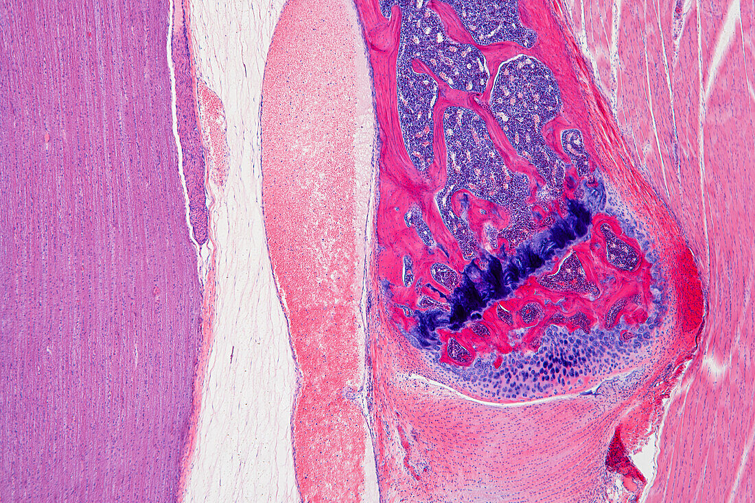
| Light micrograph of a longitudinal section through the spine of a foetus. The spinal cord, which links the brain to the rest of the body, is down left (violet). A vertebrae, one of the blocks of bone (red) that make up the spine, runs from centre right to upper centre right. Foetal bones are initially formed of cartilage (purple). Bone formation (ossification) starts from the middle of the cartilage and proceeds outwards. Areas where bone has already formed are red, areas of active ossification are dark purple. Pink and purple areas within the bone are bone marrow, a blood forming substance. An intervertebral disc (light pink), which acts as a shock absorber, can be seen beneath the vertebra. At right is muscle tissue. Magnification: x40 when printed at 15cm wide. | |
| Lizenzart: | Lizenzpflichtig |
| Credit: | Science Photo Library / EYE OF SCIENCE |
| Bildgröße: | 6016 px × 4016 px |
| Modell-Rechte: | nicht erforderlich |
| Eigentums-Rechte: | nicht erforderlich |
| Restrictions: |
|
Preise für dieses Bild ab 15 €
Universitäten & Organisationen
(Informationsmaterial Digital, Informationsmaterial Print, Lehrmaterial Digital etc.)
ab 15 €
Redaktionell
(Bücher, Bücher: Sach- und Fachliteratur, Digitale Medien (redaktionell) etc.)
ab 30 €
Werbung
(Anzeigen, Aussenwerbung, Digitale Medien, Fernsehwerbung, Karten, Werbemittel, Zeitschriften etc.)
ab 55 €
Handelsprodukte
(bedruckte Textilie, Kalender, Postkarte, Grußkarte, Verpackung etc.)
ab 75 €
Pauschalpreise
Rechtepakete für die unbeschränkte Bildnutzung in Print oder Online
ab 495 €
Keywords
- Bandscheiben,
- Biologie,
- biologisch,
- Entwicklungsbiologie,
- fötal,
- Fötus,
- gesund,
- Gewebe,
- Histologie,
- histologisch,
- Knochen,
- Knochenmark,
- lichtmikroskopische Aufnahme,
- Mikroskopie,
- Muskel,
- Nerv,
- Nervensystem,
- Niemand,
- normal,
- Ossifikation,
- Rückenmark,
- Rückgrat,
- Säule,
- Sektion,
- sektioniert,
- vertebral,
- Wirbel,
- Wirbelsäule,
- Zellen,
- zentrales Nervensystem