Retina, light micrograph
Bildnummer 13505097
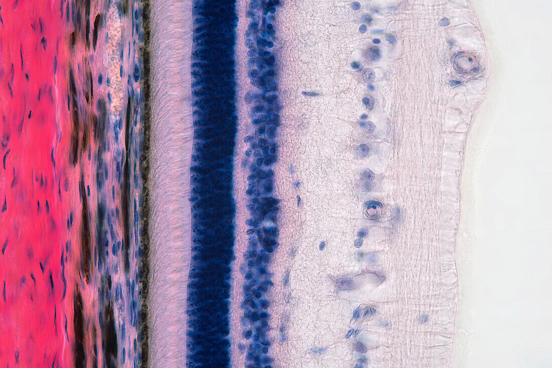
| Light micrograph of a section through the eye of the wall showing the retina. The sclera (red), the white of the eye is at left. To its right is the choroid, which contains blood vessels. Next is the retina whose first layer is a line of pigment cells (black), the retinal pigment epithelium, that lines the inside of the eye and prevents light from reflecting and distorting the image. This is immediately followed by the light sensitive rod and cone cells (light pink). For light to reach these cells it must pass through layers of nerve cells. The nerve cell nuclei are visible as the layers of purple and blue dots. Magnification: x600 when printed at 15cm wide. | |
| Lizenzart: | Lizenzpflichtig |
| Credit: | Science Photo Library / EYE OF SCIENCE |
| Bildgröße: | 6016 px × 4016 px |
| Modell-Rechte: | nicht erforderlich |
| Eigentums-Rechte: | nicht erforderlich |
| Restrictions: |
|
Preise für dieses Bild ab 15 €
Universitäten & Organisationen
(Informationsmaterial Digital, Informationsmaterial Print, Lehrmaterial Digital etc.)
ab 15 €
Redaktionell
(Bücher, Bücher: Sach- und Fachliteratur, Digitale Medien (redaktionell) etc.)
ab 30 €
Werbung
(Anzeigen, Aussenwerbung, Digitale Medien, Fernsehwerbung, Karten, Werbemittel, Zeitschriften etc.)
ab 55 €
Handelsprodukte
(bedruckte Textilie, Kalender, Postkarte, Grußkarte, Verpackung etc.)
ab 75 €
Pauschalpreise
Rechtepakete für die unbeschränkte Bildnutzung in Print oder Online
ab 495 €