Fibroblasts, fluorescent micrograph
Bildnummer 13473826
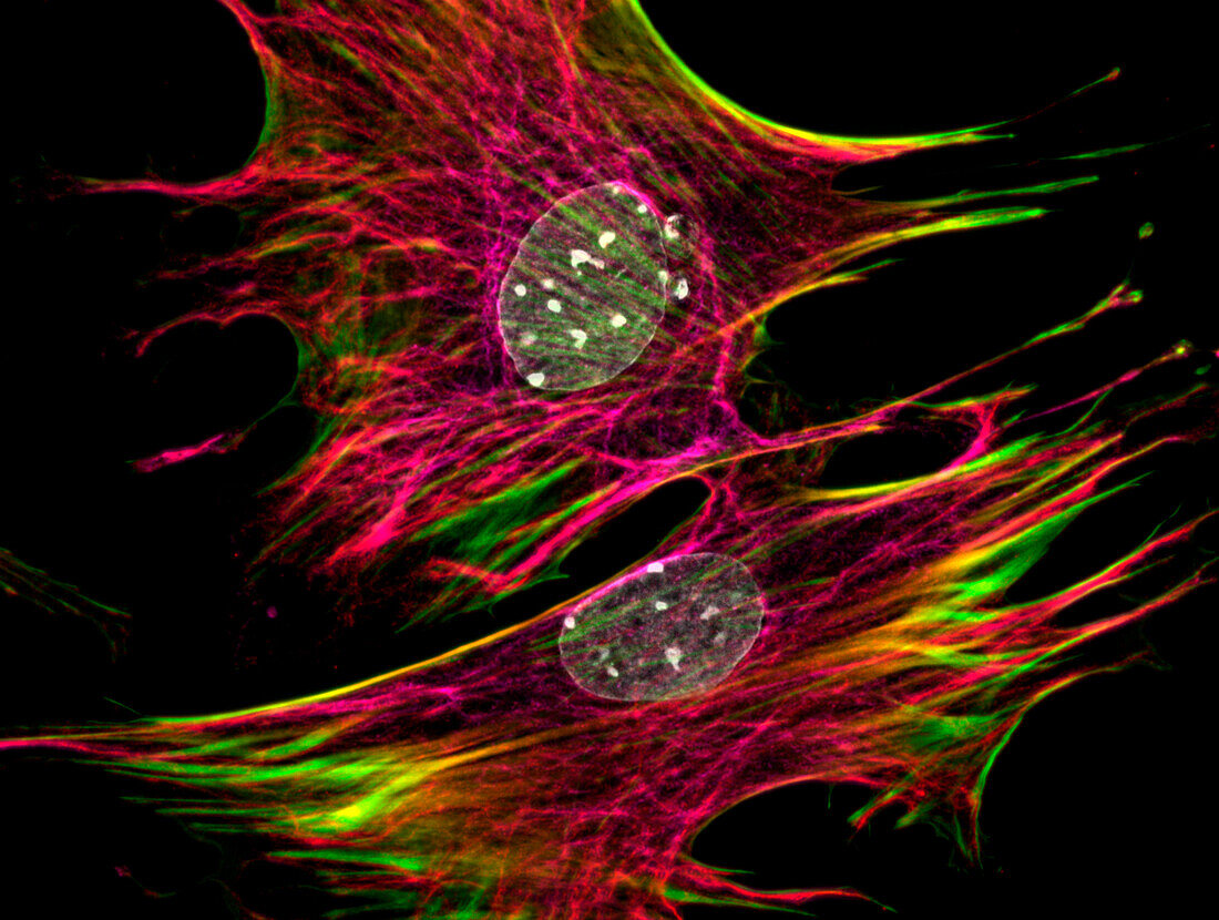
| Immunofluorescence of murine fibroblasts stained with an actin cytoskeleton probe (green/yellow), anti-FGFR-1 (fibroblast growth factor receptor 1) antibody (red/purple), and nuclear DAPI (white). Colours were achieved with multiple z-stacks. Focal planes were achieved with an optical apotome. | |
| Lizenzart: | Lizenzpflichtig |
| Credit: | Science Photo Library / JACOB C. ZBINDEN |
| Bildgröße: | 6808 px × 5134 px |
| Modell-Rechte: | nicht erforderlich |
| Eigentums-Rechte: | nicht erforderlich |
| Restrictions: | - |
Preise für dieses Bild ab 15 €
Universitäten & Organisationen
(Informationsmaterial Digital, Informationsmaterial Print, Lehrmaterial Digital etc.)
ab 15 €
Redaktionell
(Bücher, Bücher: Sach- und Fachliteratur, Digitale Medien (redaktionell) etc.)
ab 30 €
Werbung
(Anzeigen, Aussenwerbung, Digitale Medien, Fernsehwerbung, Karten, Werbemittel, Zeitschriften etc.)
ab 55 €
Handelsprodukte
(bedruckte Textilie, Kalender, Postkarte, Grußkarte, Verpackung etc.)
ab 75 €
Pauschalpreise
Rechtepakete für die unbeschränkte Bildnutzung in Print oder Online
ab 495 €