Articular cartilage and synovial membrane, light micrograph
Bildnummer 13452248
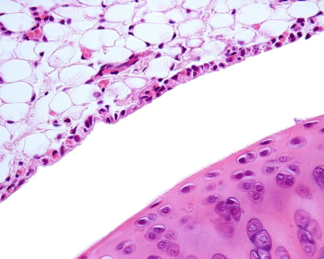
| Light micrograph of the synovial membrane delimiting the joint cavity of a diarthrosis. In the vicinity of the lumen a band of synovial cells is concentrated, adopting an appearance reminiscent of a lining epithelium, however, synovial cells are connective tissue cells that form a discontinuous lining. Above it is fatty tissue that acts like a pad on the joint. The other surface of the joint, in the lower frame, corresponds to the articular cartilage. | |
| Lizenzart: | Lizenzpflichtig |
| Credit: | Science Photo Library / JOSE CALVO |
| Bildgröße: | 3840 px × 3072 px |
| Modell-Rechte: | nicht erforderlich |
| Eigentums-Rechte: | nicht erforderlich |
| Restrictions: | - |
Preise für dieses Bild ab 15 €
Universitäten & Organisationen
(Informationsmaterial Digital, Informationsmaterial Print, Lehrmaterial Digital etc.)
ab 15 €
Redaktionell
(Bücher, Bücher: Sach- und Fachliteratur, Digitale Medien (redaktionell) etc.)
ab 30 €
Werbung
(Anzeigen, Aussenwerbung, Digitale Medien, Fernsehwerbung, Karten, Werbemittel, Zeitschriften etc.)
ab 55 €
Handelsprodukte
(bedruckte Textilie, Kalender, Postkarte, Grußkarte, Verpackung etc.)
ab 75 €
Pauschalpreise
Rechtepakete für die unbeschränkte Bildnutzung in Print oder Online
ab 495 €