Simple columnar epithelium, light micrograph
Bildnummer 13452207
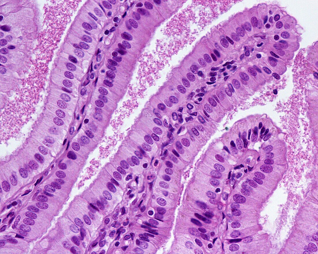
| Light micrograph of gallbladder epithelium. The gallbladder is probably the most typical example of a simple columnar epithelium. It has tall cells, with a nucleus located in the basal third, at the same height in all cells. At the apical edge there is an increase in density, little evident at this magnification, which corresponds to a border of microvilli, less developed and ordered than the striated plate of the enterocytes. Below the epithelium, constituting the axis of the folds of the mucosa, is the connective tissue of the lamina propria. | |
| Lizenzart: | Lizenzpflichtig |
| Credit: | Science Photo Library / JOSE CALVO |
| Bildgröße: | 3840 px × 3072 px |
| Modell-Rechte: | nicht erforderlich |
| Eigentums-Rechte: | nicht erforderlich |
| Restrictions: | - |
Preise für dieses Bild ab 15 €
Universitäten & Organisationen
(Informationsmaterial Digital, Informationsmaterial Print, Lehrmaterial Digital etc.)
ab 15 €
Redaktionell
(Bücher, Bücher: Sach- und Fachliteratur, Digitale Medien (redaktionell) etc.)
ab 30 €
Werbung
(Anzeigen, Aussenwerbung, Digitale Medien, Fernsehwerbung, Karten, Werbemittel, Zeitschriften etc.)
ab 55 €
Handelsprodukte
(bedruckte Textilie, Kalender, Postkarte, Grußkarte, Verpackung etc.)
ab 75 €
Pauschalpreise
Rechtepakete für die unbeschränkte Bildnutzung in Print oder Online
ab 495 €