Simple columnar epithelium, light micrograph
Bildnummer 13452206
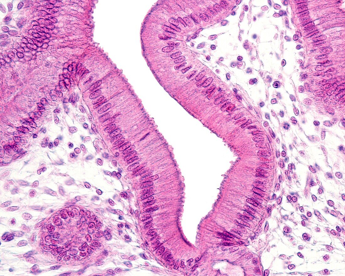
| Light micrograph of gallbladder epithelium. It is a simple columnar epithelium, with very tall cells (especially in this sample) and with very well aligned nuclei, located in the basal third. At the apical border, the cells show a density that corresponds to a border of microvilli. Lipid inclusions are frequently found in the apical cytoplasm of vesicle epithelial cells that have been removed during sample processing, leaving a small clear vacuole in their place. The connective tissue of the lamina propria is located below the epithelium, which is very loose in this sample. | |
| Lizenzart: | Lizenzpflichtig |
| Credit: | Science Photo Library / JOSE CALVO |
| Bildgröße: | 3840 px × 3072 px |
| Modell-Rechte: | nicht erforderlich |
| Eigentums-Rechte: | nicht erforderlich |
| Restrictions: | - |
Preise für dieses Bild ab 15 €
Universitäten & Organisationen
(Informationsmaterial Digital, Informationsmaterial Print, Lehrmaterial Digital etc.)
ab 15 €
Redaktionell
(Bücher, Bücher: Sach- und Fachliteratur, Digitale Medien (redaktionell) etc.)
ab 30 €
Werbung
(Anzeigen, Aussenwerbung, Digitale Medien, Fernsehwerbung, Karten, Werbemittel, Zeitschriften etc.)
ab 55 €
Handelsprodukte
(bedruckte Textilie, Kalender, Postkarte, Grußkarte, Verpackung etc.)
ab 75 €
Pauschalpreise
Rechtepakete für die unbeschränkte Bildnutzung in Print oder Online
ab 495 €