Anatomy of the colon, illustration
Bildnummer 13435261
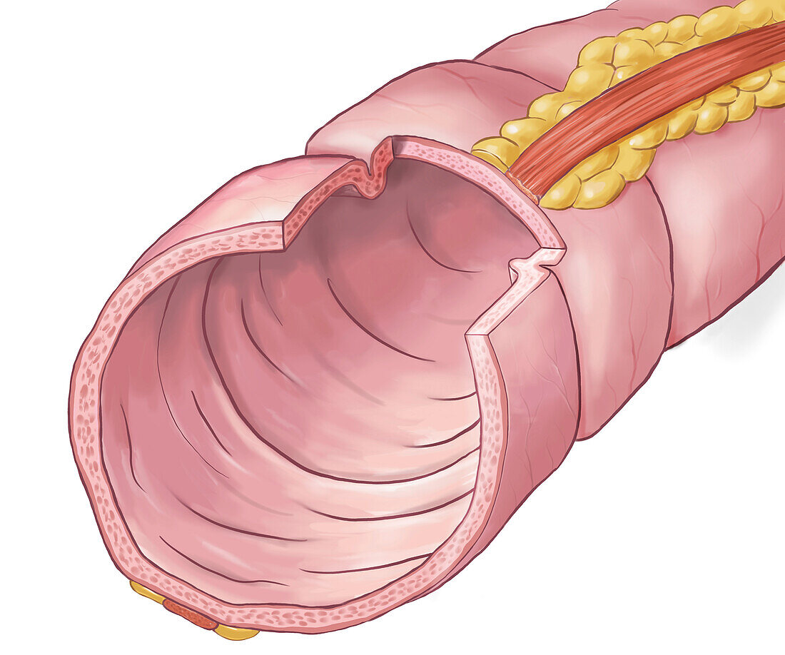
| Illustration showing a cross section of the colon. The muscular layer, plicae semilunares (folds of colon) and outpouchings, taenia libera (band of muscle, red, right) and fatty tissue (yellow) are seen. | |
| Lizenzart: | Lizenzpflichtig |
| Credit: | Science Photo Library / MICHAEL HOFFMANN / MEDICAL GRAPHICS |
| Bildgröße: | 4618 px × 3853 px |
| Modell-Rechte: | nicht erforderlich |
| Eigentums-Rechte: | nicht erforderlich |
| Restrictions: | - |
Preise für dieses Bild ab 15 €
Universitäten & Organisationen
(Informationsmaterial Digital, Informationsmaterial Print, Lehrmaterial Digital etc.)
ab 15 €
Redaktionell
(Bücher, Bücher: Sach- und Fachliteratur, Digitale Medien (redaktionell) etc.)
ab 30 €
Werbung
(Anzeigen, Aussenwerbung, Digitale Medien, Fernsehwerbung, Karten, Werbemittel, Zeitschriften etc.)
ab 55 €
Handelsprodukte
(bedruckte Textilie, Kalender, Postkarte, Grußkarte, Verpackung etc.)
ab 75 €
Pauschalpreise
Rechtepakete für die unbeschränkte Bildnutzung in Print oder Online
ab 495 €