Spinal compression, CT scan
Bildnummer 13416803
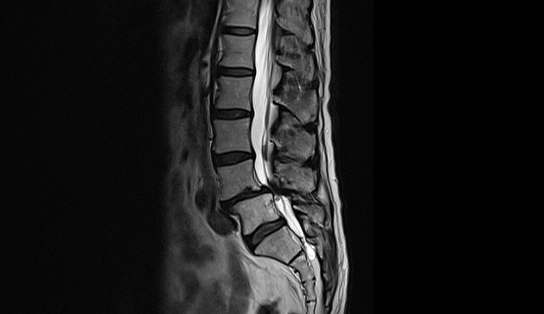
| Computed tomography (CT) scan of the lower back of an 81year old male patient with spinal compression due to ageing. The spinal cord (white) appears to be compressed (dark area) between the fourth and fifth lumbar (lower) vertebra (backbones, blocks down left). | |
| Lizenzart: | Lizenzpflichtig |
| Credit: | Science Photo Library / Marazzi, Dr. P. |
| Bildgröße: | 3516 px × 2029 px |
| Modell-Rechte: | nicht erforderlich |
| Eigentums-Rechte: | nicht erforderlich |
| Restrictions: | - |
Preise für dieses Bild ab 15 €
Universitäten & Organisationen
(Informationsmaterial Digital, Informationsmaterial Print, Lehrmaterial Digital etc.)
ab 15 €
Redaktionell
(Bücher, Bücher: Sach- und Fachliteratur, Digitale Medien (redaktionell) etc.)
ab 30 €
Werbung
(Anzeigen, Aussenwerbung, Digitale Medien, Fernsehwerbung, Karten, Werbemittel, Zeitschriften etc.)
ab 55 €
Handelsprodukte
(bedruckte Textilie, Kalender, Postkarte, Grußkarte, Verpackung etc.)
ab 75 €
Pauschalpreise
Rechtepakete für die unbeschränkte Bildnutzung in Print oder Online
ab 495 €
Keywords
- abnormal,
- achtziger Jahre,
- alternd,
- Bandscheiben,
- Computertomographie,
- CT-Scan,
- Diagnose,
- Einfarbig,
- Kompression,
- Kondition,
- Krankheit,
- L4,
- L5,
- Lendenwirbelsäule,
- Mann,
- Männlich,
- Medizin,
- medizinisch,
- menschlicher Körper,
- niedriger,
- Niemand,
- Radiographie,
- Rückenmark,
- Rückgrat,
- Scheibe,
- schwarz und weiß,
- schwarzer Hintergrund,
- Seitenansicht,
- Störung,
- ungesund,
- Wirbel,
- Wirbelsäule,
- Wirbelsäulen-,
- zentrales Nervensystem