Haematococcus pluvialis, LM
Bildnummer 13404182
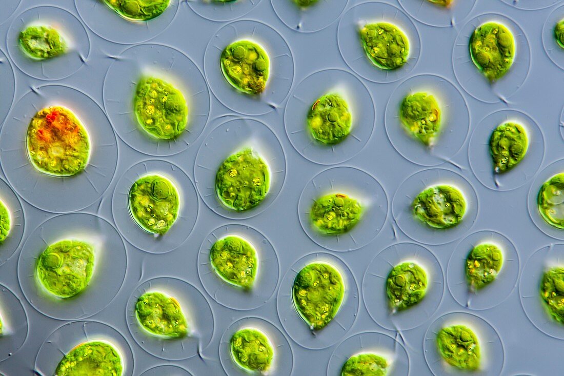
| Light micrograph of the freshwater green alga Haematococcus pluvialis. Haematococcus forms pear-shaped cells suspended in a swollen cell wall. Two flagella move the cells forward. Haematococcus is mainly found in very small waters that dry out quickly. To protect themselves from UV radiation, the cells then form a red carotenoid, astaxanthin. The water eventually turns visibly blood-red. Culture material from the algae collection at the University of Cologne (CCAC). Microscopic contrast method : Differential interference contrast. Magnification 600x at a print width of 10 cm. | |
| Lizenzart: | Lizenzpflichtig |
| Credit: | Science Photo Library / Guenther, Gerd |
| Bildgröße: | 5616 px × 3744 px |
| Modell-Rechte: | nicht erforderlich |
| Eigentums-Rechte: | nicht erforderlich |
| Restrictions: | - |
Preise für dieses Bild ab 15 €
Universitäten & Organisationen
(Informationsmaterial Digital, Informationsmaterial Print, Lehrmaterial Digital etc.)
ab 15 €
Redaktionell
(Bücher, Bücher: Sach- und Fachliteratur, Digitale Medien (redaktionell) etc.)
ab 30 €
Werbung
(Anzeigen, Aussenwerbung, Digitale Medien, Fernsehwerbung, Karten, Werbemittel, Zeitschriften etc.)
ab 55 €
Handelsprodukte
(bedruckte Textilie, Kalender, Postkarte, Grußkarte, Verpackung etc.)
ab 75 €
Pauschalpreise
Rechtepakete für die unbeschränkte Bildnutzung in Print oder Online
ab 495 €