Mixed cellularity Hodgkin lymphoma, light micrograph
Bildnummer 13387644
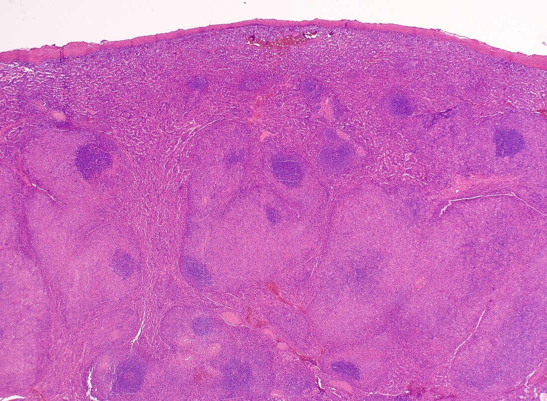
| Light micrograph of Hodgkin lymphoma, mixed cellularity subtype, involving the spleen. White pulp areas are replaced by irregular, expanded nodules. Residual lymphoid follicles are seen. Normal splenic parenchyma is seen at the top. | |
| Lizenzart: | Lizenzpflichtig |
| Credit: | Science Photo Library / WEBPATHOLOGY |
| Bildgröße: | 4096 px × 3000 px |
| Modell-Rechte: | nicht erforderlich |
| Eigentums-Rechte: | nicht erforderlich |
| Restrictions: | - |
Preise für dieses Bild ab 15 €
Universitäten & Organisationen
(Informationsmaterial Digital, Informationsmaterial Print, Lehrmaterial Digital etc.)
ab 15 €
Redaktionell
(Bücher, Bücher: Sach- und Fachliteratur, Digitale Medien (redaktionell) etc.)
ab 30 €
Werbung
(Anzeigen, Aussenwerbung, Digitale Medien, Fernsehwerbung, Karten, Werbemittel, Zeitschriften etc.)
ab 55 €
Handelsprodukte
(bedruckte Textilie, Kalender, Postkarte, Grußkarte, Verpackung etc.)
ab 75 €
Pauschalpreise
Rechtepakete für die unbeschränkte Bildnutzung in Print oder Online
ab 495 €
Keywords
- Biologie,
- biologisch,
- CD15,
- EBV,
- Hämatologie,
- hämatologisch,
- Hämatopathologie,
- Histologie,
- histologisch,
- Histopathologie,
- histopathologisch,
- Hodgkin-Lymphom,
- Hodgkin-Zelle,
- Hodgkinsche Krankheit,
- Krebs,
- krebsartig,
- lakunäre Zelle,
- lichtmikroskopische Aufnahme,
- LP-Zelle,
- Lymphknoten,
- maligne,
- Malignom,
- Mikroskopie,
- Neoplasma,
- Niemand,
- Onkologie,
- Pathologie,
- Popcorn-Zelle,
- Zentrum