Eye anatomy, illustration
Bildnummer 13376764
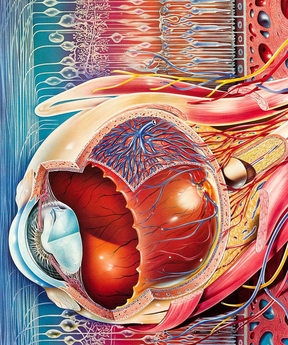
| Eye anatomy. Cutaway illustration of the eye, from the side, with the structure of the retina in the background. Structures shown include the lens (light blue, lower left), its attachment points, the iris, and the bulge of the cornea at the front of the eye. Several layers of the eyeball are shown, including the sclera (outer layer), the choroid, and the retina (innermost). Retinal capillaries (red and blue) are shown. The optic nerve (yellow) is at far right. External structures shown include muscles (red) that control the movement of the eyeball, and nerves (yellow) and more arteries (red) and veins (blue). | |
| Lizenzart: | Lizenzpflichtig |
| Credit: | Science Photo Library / Bavosi, John |
| Bildgröße: | 4620 px × 5526 px |
| Modell-Rechte: | nicht erforderlich |
| Eigentums-Rechte: | nicht erforderlich |
| Restrictions: | - |
Preise für dieses Bild ab 15 €
Universitäten & Organisationen
(Informationsmaterial Digital, Informationsmaterial Print, Lehrmaterial Digital etc.)
ab 15 €
Redaktionell
(Bücher, Bücher: Sach- und Fachliteratur, Digitale Medien (redaktionell) etc.)
ab 30 €
Werbung
(Anzeigen, Aussenwerbung, Digitale Medien, Fernsehwerbung, Karten, Werbemittel, Zeitschriften etc.)
ab 55 €
Handelsprodukte
(bedruckte Textilie, Kalender, Postkarte, Grußkarte, Verpackung etc.)
ab 75 €
Pauschalpreise
Rechtepakete für die unbeschränkte Bildnutzung in Print oder Online
ab 495 €
Keywords
- Aderhaut,
- Anatomie,
- anatomisch,
- Arterie,
- arteriell,
- Arterien,
- aufgegliedert,
- Augapfel,
- Auge,
- Augen-,
- Augenheilkunde,
- Biologie,
- biologisch,
- Blutgefäße,
- Cutaway,
- gesund,
- Gewebe,
- Hornhaut,
- Illustration,
- Iris,
- Kapillarbett,
- Kunstwerk,
- Linse,
- menschlicher Körper,
- Muskeln,
- Nerven,
- Netzhaut-,
- Neurologie,
- neurologisch,
- Niemand,
- normal,
- okular,
- Organ,
- Retina,
- sensorisch,
- Sicht,
- Sinn,
- vaskulär,
- Vene,
- Venen,
- venös,
- Vision,
- visuell