Infected tick salivary gland, light micrograph
Bildnummer 13369203
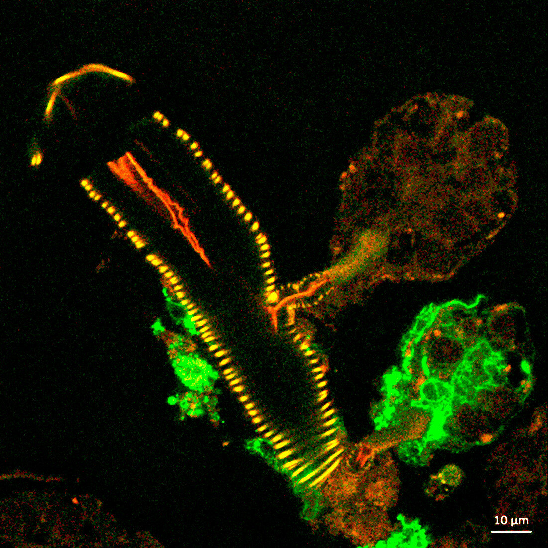
| Infected tick salivary gland. Confocal light micrograph of a cross-section of a tick salivary gland infected with Langat virus (green), a flavivirus with a positive-sense RNA (ribonucleic acid) genome. The two rounded structures on the right, acini, are shown attached to a duct (yellow). The lower acinus is infected, as shown by the green fluorescent signal. US National Institutes of Health (NIH) scientists are exploring black-legged tick (Ixodes scapularis) salivary glands as a tool for studying flavivirus transmission and infection. | |
| Lizenzart: | Lizenzpflichtig |
| Credit: | Science Photo Library / NATIONAL INSTITUTES OF HEALTH |
| Bildgröße: | 3000 px × 3000 px |
| Modell-Rechte: | nicht erforderlich |
| Eigentums-Rechte: | nicht erforderlich |
| Restrictions: | - |
Preise für dieses Bild ab 15 €
Universitäten & Organisationen
(Informationsmaterial Digital, Informationsmaterial Print, Lehrmaterial Digital etc.)
ab 15 €
Redaktionell
(Bücher, Bücher: Sach- und Fachliteratur, Digitale Medien (redaktionell) etc.)
ab 30 €
Werbung
(Anzeigen, Aussenwerbung, Digitale Medien, Fernsehwerbung, Karten, Werbemittel, Zeitschriften etc.)
ab 55 €
Handelsprodukte
(bedruckte Textilie, Kalender, Postkarte, Grußkarte, Verpackung etc.)
ab 75 €
Pauschalpreise
Rechtepakete für die unbeschränkte Bildnutzung in Print oder Online
ab 495 €
Keywords
- ansteckend,
- Biologie,
- biologisch,
- Erreger,
- Flavivirus,
- Fluoreszenz,
- fluoreszierend,
- Forschung,
- Gang,
- GFP,
- Grün fluoreszierendes Protein,
- Infektion,
- infiziert,
- Ixodes,
- Kondition,
- konfokale Lichtmikroskopie,
- konfokale mikroskopische Aufnahme,
- Krankheit,
- Mikroskopie,
- Niemand,
- pathogen,
- RNA-Virus,
- schwarzer Hintergrund,
- Störung,
- Tick,
- Übertragung,
- Vektor,
- Virologie,
- virologisch,
- Virus