Female reproductive organs and pelvic bones,CT-based image
Bildnummer 12967559
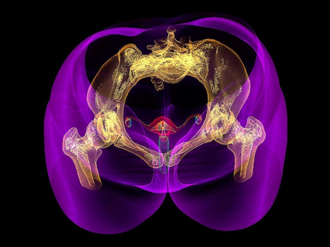
| Female reproductive organs and pelvic bones,3D image based on computed tomography (CT) scan. The pelvic and hip bones are yellow in this semi-transparent view from above. At centre is the uterus,within which a foetus develops during pregnancy. Below it is the vagina,which also functions as the birth canal. Between the two is the cervix,the neck of the uterus. At left and right are the two ovaries,from one of which an egg (ovum) is released during the monthly ovulation cycle. This egg travels down the adjacent oviduct (Fallopian tube) to the uterus,where it either embeds in the uterine wall (if fertilised) or is lost with the uterine wall (endometrium) during menstruation. | |
| Lizenzart: | Lizenzpflichtig |
| Credit: | Science Photo Library / Fung, K.H. |
| Bildgröße: | 5294 px × 3962 px |
| Modell-Rechte: | nicht erforderlich |
| Eigentums-Rechte: | nicht erforderlich |
| Restrictions: | - |
Preise für dieses Bild ab 15 €
Universitäten & Organisationen
(Informationsmaterial Digital, Informationsmaterial Print, Lehrmaterial Digital etc.)
ab 15 €
Redaktionell
(Bücher, Bücher: Sach- und Fachliteratur, Digitale Medien (redaktionell) etc.)
ab 30 €
Werbung
(Anzeigen, Aussenwerbung, Digitale Medien, Fernsehwerbung, Karten, Werbemittel, Zeitschriften etc.)
ab 55 €
Handelsprodukte
(bedruckte Textilie, Kalender, Postkarte, Grußkarte, Verpackung etc.)
ab 75 €
Pauschalpreise
Rechtepakete für die unbeschränkte Bildnutzung in Print oder Online
ab 495 €
Keywords
- Abdomen,
- Anatomie,
- anatomisch,
- anterior,
- ausgeschnitten,
- Ausschnitte,
- Bänder,
- Bauch,
- Biologie,
- biologisch,
- Computertomographie,
- CT-Scan,
- Eierstock,
- Eierstock-,
- Eierstöcke,
- Eileiter,
- farbig,
- Fortpflanzungssystem,
- Frontal,
- Gebärmutter,
- Gebärmutterhals,
- Gebärmutterhals-,
- gefärbt,
- gesund,
- Hüften,
- Illustration,
- Knochen,
- Kunstwerk,
- menschlicher Körper,
- Niemand,
- normal,
- Organ,
- Organe,
- pelvin,
- Reproduktion,
- Scanner,
- schwarzer Hintergrund,
- uterin,
- Uterus,
- Vagina,
- vaginal,
- Vorderseite,
- Weiblich,
- weibliche Fortpflanzungsorgane,
- weibliches Fortpflanzungssystem