Normal heart,ultrasound scan
Bildnummer 12960857
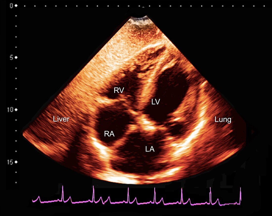
| Normal heart. Coloured ultrasound scan of a healthy heart in a 49-year-old man during the systolic phase of the heartbeat. This scan passes vertically through the heart,with the heart's four chambers seen as the black areas. The central coloured areas are the walls and valves separating the chambers. The top of the heart is at bottom. The top two chambers are the ventricles (RV and LV),and the bottom two chambers are the atria (RA and LA). The liver and a lung are also labelled. The heart is a hollow muscular pump that contracts continuously under a steady cycle of electrical impulses,sending blood to the lungs and around the body. An electrocardiogram (ECG) trace is seen across bottom,with the peaks of electrical activity corresponding to the beating of the heart. Ultrasound scanning uses high-frequency sound waves to look at structures inside the body. | |
| Lizenzart: | Lizenzpflichtig |
| Credit: | Science Photo Library / Zephyr |
| Bildgröße: | 4293 px × 3402 px |
| Modell-Rechte: | nicht erforderlich |
| Eigentums-Rechte: | nicht erforderlich |
| Restrictions: | - |
Preise für dieses Bild ab 15 €
Universitäten & Organisationen
(Informationsmaterial Digital, Informationsmaterial Print, Lehrmaterial Digital etc.)
ab 15 €
Redaktionell
(Bücher, Bücher: Sach- und Fachliteratur, Digitale Medien (redaktionell) etc.)
ab 30 €
Werbung
(Anzeigen, Aussenwerbung, Digitale Medien, Fernsehwerbung, Karten, Werbemittel, Zeitschriften etc.)
ab 55 €
Handelsprodukte
(bedruckte Textilie, Kalender, Postkarte, Grußkarte, Verpackung etc.)
ab 75 €
Pauschalpreise
Rechtepakete für die unbeschränkte Bildnutzung in Print oder Online
ab 495 €
Keywords
- 40er Jahre,
- Anatomie,
- anatomisch,
- Atrium,
- beschriftet,
- Diagnose,
- Echokardiogramm,
- Elektrokardiographie,
- Erwachsene,
- Etikette,
- Etiketten,
- farbig,
- gefärbt,
- Gefäßsystem,
- gesund,
- Herz,
- Kardiologie,
- kardiovaskular,
- Kreislauf,
- Leber,
- links,
- Lungen,
- Mann,
- Männlich,
- menschlicher Körper,
- Muskel,
- Niemand,
- normal,
- Organ,
- Physiologie,
- physiologisch,
- Recht,
- Scanner,
- schwarzer Hintergrund,
- Sonographie,
- Spur,
- Ultraschalluntersuchung,
- Ventrikel,
- Vierziger Jahre