Lung cancer SEM-TEM comparison
Bildnummer 12948540
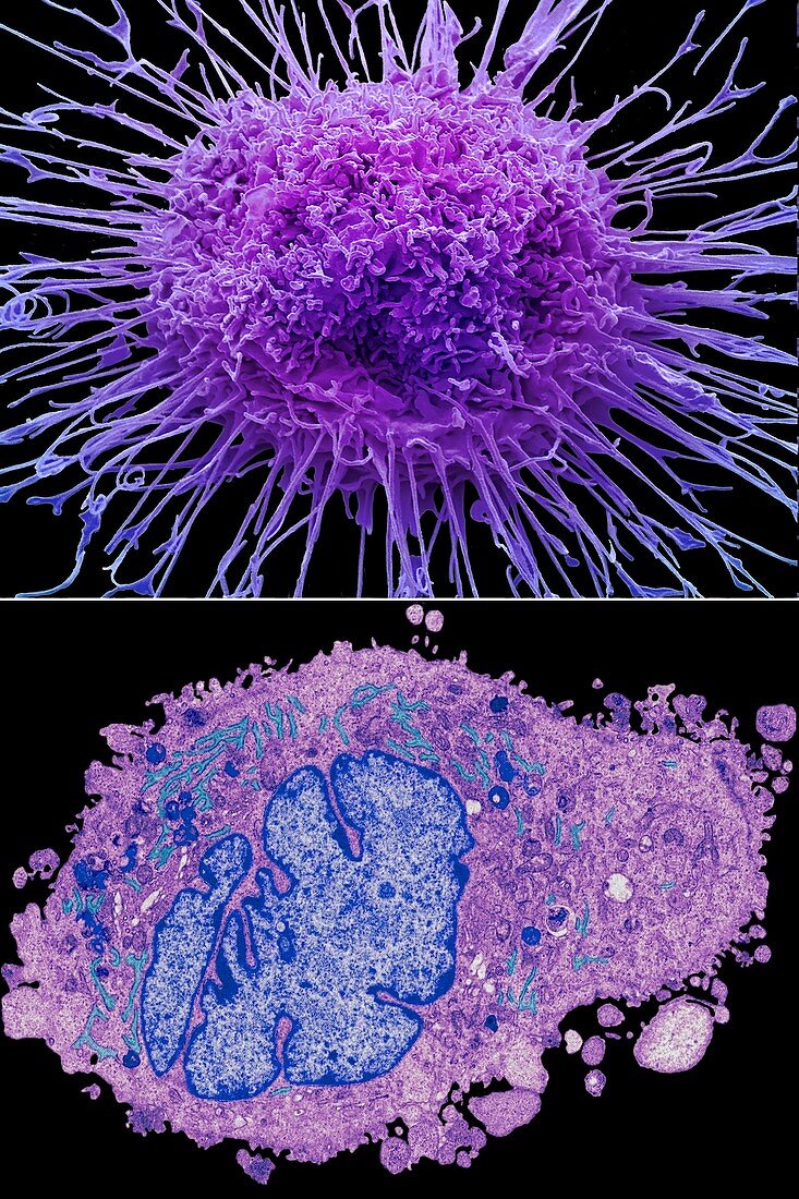
| Comparison between a scanning electron micrograph (SEM , top) and transmission electron micrograph (TEM, bottom) of a lung cancer cell. Typical of many cancer cells the nucleus (blue in TEM) is enlarged and multilobed, the cell walls are covered in projections (SEM) and the cytoplasm contains a large number of organelles (TEM). Adenocarcinoma (shown here) is a non-small cell lung cancer that is more commonly found in women and non-smokers, and it is the most common type of lung cancer for people under 45. Adenocarcinoma accounts for approximately 50% of all non-small cell lung cancers and begins in the outer sections of the lung (although it can occur as central lesions) which can make it difficult to detect in the early stages of the disease. Magnification: x4000 at 10 centimetres wide. For a series of comparisons between SEMs and TEMs see images C047/7006 to C047/7034. | |
| Lizenzart: | Lizenzpflichtig |
| Credit: | Science Photo Library / Gschmeissner, Steve |
| Bildgröße: | 4572 px × 6858 px |
| Modell-Rechte: | nicht erforderlich |
| Eigentums-Rechte: | nicht erforderlich |
| Restrictions: | - |
Preise für dieses Bild ab 15 €
Universitäten & Organisationen
(Informationsmaterial Digital, Informationsmaterial Print, Lehrmaterial Digital etc.)
ab 15 €
Redaktionell
(Bücher, Bücher: Sach- und Fachliteratur, Digitale Medien (redaktionell) etc.)
ab 30 €
Werbung
(Anzeigen, Aussenwerbung, Digitale Medien, Fernsehwerbung, Karten, Werbemittel, Zeitschriften etc.)
ab 55 €
Handelsprodukte
(bedruckte Textilie, Kalender, Postkarte, Grußkarte, Verpackung etc.)
ab 75 €
Pauschalpreise
Rechtepakete für die unbeschränkte Bildnutzung in Print oder Online
ab 495 €
Keywords
- abnormal,
- Atomkern,
- farbig,
- gefärbt,
- Gesundheitswesen,
- Kondition,
- Krankheit,
- Krebs,
- krebsartig,
- Lunge,
- maligne,
- Malignom,
- Medizin,
- medizinisch,
- Mikrovilli,
- Organellen,
- rasterelektronenmikroskopische Aufnahme,
- Reihenfolge,
- REM,
- Serie,
- Störung,
- System,
- tem,
- transmissionselektronenmikroskopische Aufnahme,
- Tumor,
- Tumoren,
- Vergleich,
- vergleichen,
- verglichen,
- Wachstum,
- Zelle,
- Zellen