Frontal sinus infection,CT scan
Bildnummer 12894704
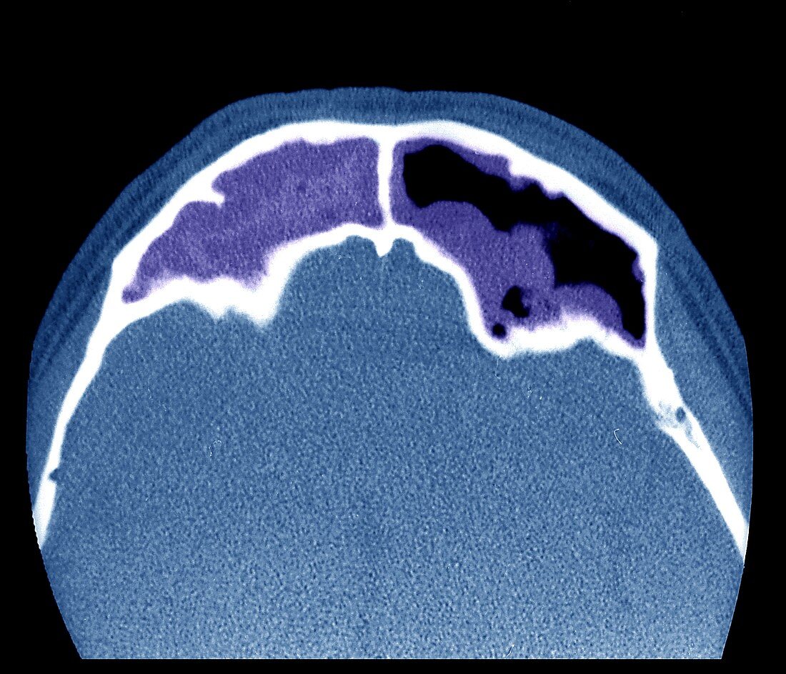
| Frontal sinus infection. Coloured computerized tomography (CT) scan of an axial section of the brain of a 32-year-old patient with a frontal sinus infection. It is a bone window study (grey-level mapping,involving manipulation of the greyscale component of a CT scan to highlight specific structures in the brain),centred on the frontal sinuses. There is an infection of the right frontal sinus,along with a partial filling of the left frontal sinus. The frontal (paranasal) sinuses are airspaces (lined with a mucosal membrane) within the bones of the face and skull. Sinus infection (sinusitis) is where these airspaces become filled with fluid,which leads to bacterial growth and viruses causing infection. This patient is prone to a global sinus infection. This is a CT scan without intravenous iodine injection. | |
| Lizenzart: | Lizenzpflichtig |
| Credit: | Science Photo Library / Zephyr |
| Bildgröße: | 3858 px × 3307 px |
| Modell-Rechte: | nicht erforderlich |
| Eigentums-Rechte: | nicht erforderlich |
| Restrictions: | - |
Preise für dieses Bild ab 15 €
Universitäten & Organisationen
(Informationsmaterial Digital, Informationsmaterial Print, Lehrmaterial Digital etc.)
ab 15 €
Redaktionell
(Bücher, Bücher: Sach- und Fachliteratur, Digitale Medien (redaktionell) etc.)
ab 30 €
Werbung
(Anzeigen, Aussenwerbung, Digitale Medien, Fernsehwerbung, Karten, Werbemittel, Zeitschriften etc.)
ab 55 €
Handelsprodukte
(bedruckte Textilie, Kalender, Postkarte, Grußkarte, Verpackung etc.)
ab 75 €
Pauschalpreise
Rechtepakete für die unbeschränkte Bildnutzung in Print oder Online
ab 495 €
Keywords
- 30er Jahre,
- abnormal,
- axial,
- Axialschnitt,
- bakteriell,
- bakterielle Infektion,
- Bakterien,
- Bakterium,
- Computertomographie,
- ct,
- CT-Scan,
- dreißiger Jahre,
- farbig,
- Fluid,
- Füllung,
- geduldig,
- gefärbt,
- Gehirn,
- Hirnscan,
- Infektion,
- klinisch,
- Kondition,
- Krankheit,
- Luftraum,
- Medizin,
- medizinisch,
- menschlicher Körper,
- Mikroorganismus,
- Nasennebenhöhlen,
- Nebenhöhlen,
- Nebenhöhlenentzündung,
- Neurologie,
- neurologisch,
- Neurowissenschaften,
- Niemand,
- Radiologie,
- radiologisch,
- Scan,
- Scanner,
- Schädel,
- schwarzer Hintergrund,
- Sinus,
- Stirnhöhlen,
- Störung,
- ungesund,
- viral,
- Viren,
- Virus