Gene profiling for predicting colon cancer
Bildnummer 12652637
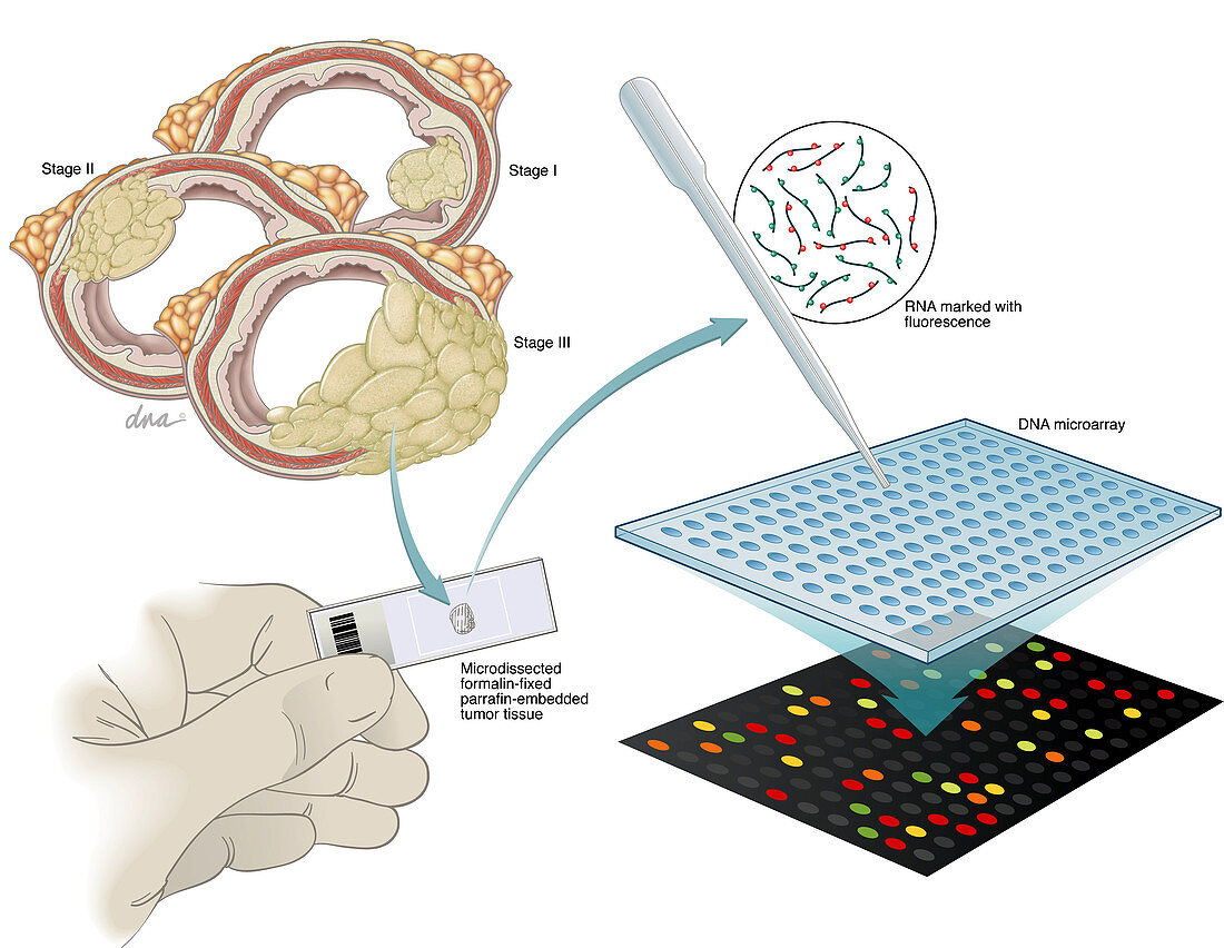
| This illustration shows three cross-sections of the large intestine showing stages 1 to 3 of colon cancer, which was used in development studies. The RNA is extracted from a microdissection of the tumour tissue that has been fixed with formalin and embedded in paraffin. The RNA genes are marked with fluorescence and applied to a DNA microarray. Seven genes were used in the assay to determine risk of recurrence. | |
| Lizenzart: | Lizenzpflichtig |
| Credit: | Science Photo Library / Science Source / DNA Illustrations |
| Bildgröße: | 3358 px × 2601 px |
| Modell-Rechte: | nicht erforderlich |
| Eigentums-Rechte: | nicht erforderlich |
| Restrictions: | - |
Preise für dieses Bild ab 15 €
Universitäten & Organisationen
(Informationsmaterial Digital, Informationsmaterial Print, Lehrmaterial Digital etc.)
ab 15 €
Redaktionell
(Bücher, Bücher: Sach- und Fachliteratur, Digitale Medien (redaktionell) etc.)
ab 30 €
Werbung
(Anzeigen, Aussenwerbung, Digitale Medien, Fernsehwerbung, Karten, Werbemittel, Zeitschriften etc.)
ab 55 €
Handelsprodukte
(bedruckte Textilie, Kalender, Postkarte, Grußkarte, Verpackung etc.)
ab 75 €
Pauschalpreise
Rechtepakete für die unbeschränkte Bildnutzung in Print oder Online
ab 495 €