Slipped disc in the lumbar spine, MRI scan
Bildnummer 12644504
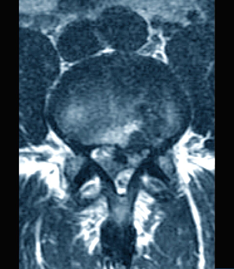
| Slipped disc in the lumbar spine. Axial magnetic resonance imaging (MRI) scan through a slipped (herniated) intervertebral disc in the lumbar spine of a 39-year-old man. This herniation is of the disc between the L4 and L5 vertebrae, causing a bulge (white area at lower centre) outwards putting pressure on the spinal cord. This pressure can cause severe pain and other neurological symptoms. In this case, it has caused paralytic sciatica (paralysis and pain shooting down the leg) and sphincter disorders (affecting bowel movements). This is a severe herniation of the disc, with a disc fragment having migrated outwards. For a series of images of this condition, including arrows showing the location of the disc bulge, see C040/3347 to C040/3354. | |
| Lizenzart: | Lizenzpflichtig |
| Credit: | Science Photo Library / Zephyr |
| Bildgröße: | 3904 px × 4476 px |
| Modell-Rechte: | nicht erforderlich |
| Eigentums-Rechte: | nicht erforderlich |
| Restrictions: | - |
Preise für dieses Bild ab 15 €
Universitäten & Organisationen
(Informationsmaterial Digital, Informationsmaterial Print, Lehrmaterial Digital etc.)
ab 15 €
Redaktionell
(Bücher, Bücher: Sach- und Fachliteratur, Digitale Medien (redaktionell) etc.)
ab 30 €
Werbung
(Anzeigen, Aussenwerbung, Digitale Medien, Fernsehwerbung, Karten, Werbemittel, Zeitschriften etc.)
ab 55 €
Handelsprodukte
(bedruckte Textilie, Kalender, Postkarte, Grußkarte, Verpackung etc.)
ab 75 €
Pauschalpreise
Rechtepakete für die unbeschränkte Bildnutzung in Print oder Online
ab 495 €
Keywords
- 30er Jahre,
- abnormal,
- Arthrologie,
- axial,
- Bandscheiben,
- Blau,
- chirurgisch,
- dreißiger Jahre,
- Einfarbig,
- eingeklemmter Nerv,
- Erwachsene,
- geduldig,
- Gelenk,
- Joint,
- Kondition,
- Krankheit,
- L4,
- L5,
- Lendenwirbelsäule,
- Magnetresonanztomografie,
- Mann,
- Männlich,
- Medizin,
- medizinisch,
- menschlicher Körper,
- MRT-Untersuchung,
- Neurologie,
- neurologisch,
- Niemand,
- Querschnitt,
- Rücken,
- Rückgrat,
- Scanner,
- Schließmuskel,
- Sektion,
- sektioniert,
- ungesund,
- Verletzung,
- Wirbelsäule