Compression fracture of a lumbar vertebra, X-ray
Bildnummer 12644485
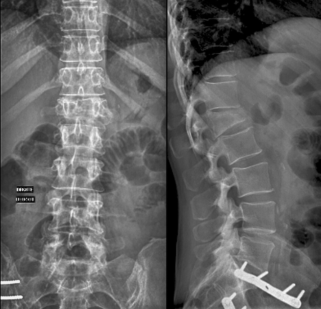
| Compression fracture of a lumbar vertebra. Frontal (left) and lateral (right) X-rays of the vertebral column of a 47-year-old woman with multiple injuries. The compression fracture is to the L1 vertebra, which is at upper centre. Due to its collapse, this vertebra is smaller and appears wedge-shaped in the lateral view when compared to the normal surrounding vertebrae. | |
| Lizenzart: | Lizenzpflichtig |
| Credit: | Science Photo Library / Zephyr |
| Bildgröße: | 4258 px × 4104 px |
| Modell-Rechte: | nicht erforderlich |
| Eigentums-Rechte: | nicht erforderlich |
| Restrictions: | - |
Preise für dieses Bild ab 15 €
Universitäten & Organisationen
(Informationsmaterial Digital, Informationsmaterial Print, Lehrmaterial Digital etc.)
ab 15 €
Redaktionell
(Bücher, Bücher: Sach- und Fachliteratur, Digitale Medien (redaktionell) etc.)
ab 30 €
Werbung
(Anzeigen, Aussenwerbung, Digitale Medien, Fernsehwerbung, Karten, Werbemittel, Zeitschriften etc.)
ab 55 €
Handelsprodukte
(bedruckte Textilie, Kalender, Postkarte, Grußkarte, Verpackung etc.)
ab 75 €
Pauschalpreise
Rechtepakete für die unbeschränkte Bildnutzung in Print oder Online
ab 495 €
Keywords
- 40er Jahre,
- abnormal,
- anterior,
- Arthrologie,
- Einfarbig,
- Erwachsene,
- Frau,
- Frontal,
- geduldig,
- Knochen,
- Kompression,
- L1,
- Lendenwirbelsäule,
- Medizin,
- medizinisch,
- menschlicher Körper,
- Niemand,
- Osteologie,
- Profil,
- Radiographie,
- Röntgen,
- Röntgengerät,
- Rücken,
- Rückgrat,
- Schwarz und weiß,
- Seitenansicht,
- seitlich,
- thorakal,
- Unfall,
- ungesund,
- verletzt,
- Verletzung,
- vertebral,
- Vierziger Jahre,
- Vorderansicht,
- Weiblich,
- Wirbelsäule,
- Wirbelsäulen-