Brain arteries and venous sinuses, 3D CT angiogram
Bildnummer 12644404
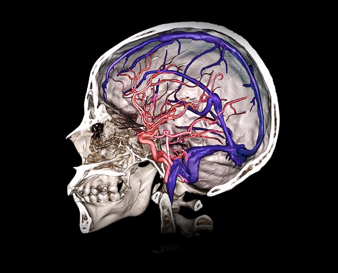
| Brain arteries and venous sinuses. Coloured lateral 3D computed tomography (CT) angiogram, showing a cutaway view of the skull and some of the brain arteries (pink) and venous sinuses (purple) of a 35-year-old man. The major internal neck arteries (carotid and vertebral) are shown meeting at the circle of Willis and branching out into the brain. The venous sinuses receive deoxygenated blood from the brain's veins and mostly channel it to the internal jugular veins in the neck. | |
| Lizenzart: | Lizenzpflichtig |
| Credit: | Science Photo Library / Zephyr |
| Bildgröße: | 4648 px × 3759 px |
| Modell-Rechte: | nicht erforderlich |
| Eigentums-Rechte: | nicht erforderlich |
| Restrictions: | - |
Preise für dieses Bild ab 15 €
Universitäten & Organisationen
(Informationsmaterial Digital, Informationsmaterial Print, Lehrmaterial Digital etc.)
ab 15 €
Redaktionell
(Bücher, Bücher: Sach- und Fachliteratur, Digitale Medien (redaktionell) etc.)
ab 30 €
Werbung
(Anzeigen, Aussenwerbung, Digitale Medien, Fernsehwerbung, Karten, Werbemittel, Zeitschriften etc.)
ab 55 €
Handelsprodukte
(bedruckte Textilie, Kalender, Postkarte, Grußkarte, Verpackung etc.)
ab 75 €
Pauschalpreise
Rechtepakete für die unbeschränkte Bildnutzung in Print oder Online
ab 495 €
Keywords
- 3 dimensional,
- 3-d,
- 3-dimensional,
- 30er Jahre,
- 3D,
- Anatomie,
- anatomisch,
- Angiografie,
- Angiogramm,
- Arterie,
- arteriell,
- Arterien,
- ausgeschnitten,
- Ausschnitte,
- Biologie,
- biologisch,
- Blutgefäß,
- Computertomographie,
- CT-Scan,
- Cutaway,
- Dreidimensional,
- dreißiger Jahre,
- Erwachsene,
- farbig,
- gefärbt,
- Gehirn,
- gesund,
- Kopf,
- kranial,
- Kreislauf,
- Mann,
- Männlich,
- menschlicher Körper,
- Niemand,
- normal,
- Profil,
- Radiographie,
- Röntgen,
- Scanner,
- Schädel,
- schwarzer Hintergrund,
- Seitenansicht,
- seitlich,
- vaskulär,
- Venen,
- vertebral