Red and white muscle fibres, TEM
Bildnummer 12582307
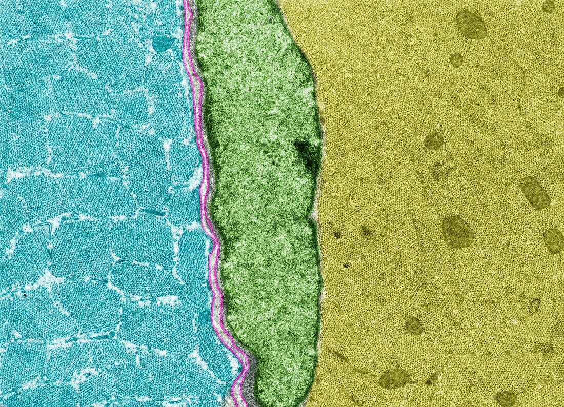
| Coloured transmission electron micrograph (TEM) showing cross-sectioned white (blue) and red (yellow) muscle fibres. The white fibre shows more cytoplasm between the myofibrils. Also seen is a cell nucleus (green) and basal lamina (pink). | |
| Lizenzart: | Lizenzpflichtig |
| Credit: | Science Photo Library / Jose Calvo |
| Bildgröße: | 4919 px × 3552 px |
| Modell-Rechte: | nicht erforderlich |
| Eigentums-Rechte: | nicht erforderlich |
| Restrictions: | - |
Preise für dieses Bild ab 15 €
Universitäten & Organisationen
(Informationsmaterial Digital, Informationsmaterial Print, Lehrmaterial Digital etc.)
ab 15 €
Redaktionell
(Bücher, Bücher: Sach- und Fachliteratur, Digitale Medien (redaktionell) etc.)
ab 30 €
Werbung
(Anzeigen, Aussenwerbung, Digitale Medien, Fernsehwerbung, Karten, Werbemittel, Zeitschriften etc.)
ab 55 €
Handelsprodukte
(bedruckte Textilie, Kalender, Postkarte, Grußkarte, Verpackung etc.)
ab 75 €
Pauschalpreise
Rechtepakete für die unbeschränkte Bildnutzung in Print oder Online
ab 495 €