Small intestine lining, SEM
Bildnummer 12528278
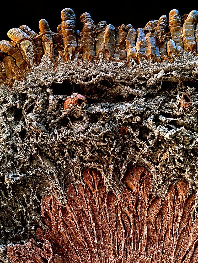
| Small intestine lining. Coloured scanning electron micrograph (SEM) of a section through the mucosa (lining) of a small intestine. Villi (brown, across top) project into the intestine lumen (cavity), greatly increasing the surface area for the absorption of nutrients from food. Below the villi is the submucosa (grey). This tissue contains capillary blood vessels (red, for example at upper right), which transport absorbed nutrients to a nearby vein. The submucosa also contains lymph vessels and fat cells (not seen). Beneath the submucosa is the muscularis mucosae (red, bottom), a layer of muscle tissue. Magnification: x24 at 6x7cm size. x40 at 4x5ins | |
| Lizenzart: | Lizenzpflichtig |
| Credit: | Science Photo Library / EYE OF SCIENCE |
| Bildgröße: | 3669 px × 4866 px |
| Modell-Rechte: | nicht erforderlich |
| Eigentums-Rechte: | nicht erforderlich |
| Restrictions: |
|
Preise für dieses Bild ab 15 €
Universitäten & Organisationen
(Informationsmaterial Digital, Informationsmaterial Print, Lehrmaterial Digital etc.)
ab 15 €
Redaktionell
(Bücher, Bücher: Sach- und Fachliteratur, Digitale Medien (redaktionell) etc.)
ab 30 €
Werbung
(Anzeigen, Aussenwerbung, Digitale Medien, Fernsehwerbung, Karten, Werbemittel, Zeitschriften etc.)
ab 55 €
Handelsprodukte
(bedruckte Textilie, Kalender, Postkarte, Grußkarte, Verpackung etc.)
ab 75 €
Pauschalpreise
Rechtepakete für die unbeschränkte Bildnutzung in Print oder Online
ab 495 €
Keywords
- Absorption,
- Anatomie,
- Blutgefäß,
- Darm,
- Dünndarm,
- farbig,
- Futter,
- Gefäße,
- gesund,
- Gewebe,
- kapillar,
- Kreislauf,
- Mauer,
- menschlicher Körper,
- Muskel,
- Muskulös,
- normal,
- Projektion,
- Projektionen,
- rasterelektronenmikroskopische Aufnahme,
- REM,
- Schleimhaut,
- sekretorisch,
- Sektion,
- sektioniert,
- Submukosa,
- System,
- Trakt,
- vaskulär,
- Verdauung,
- Verdauungskanal,
- Verdauungssystem,
- Zotte,
- Zotten