Glial limiting membrane, TEM
Bildnummer 12504725
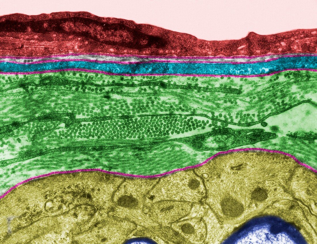
| Coloured transmission electron micrograph (TEM) showing the glial limiting membrane (glia limitans) of white matter. It is formed by astrocyte processes (yellow) that separated blood capillary endothelium (red), pericyte (blue) and the surrounding connective tissue (green) from the myelinated nerve fibres (dark purple, bottom). Basal lamina is pink. | |
| Lizenzart: | Lizenzpflichtig |
| Credit: | Science Photo Library / JOSE CALVO |
| Bildgröße: | 3622 px × 2790 px |
| Modell-Rechte: | nicht erforderlich |
| Eigentums-Rechte: | nicht erforderlich |
| Restrictions: | - |
Preise für dieses Bild ab 15 €
Universitäten & Organisationen
(Informationsmaterial Digital, Informationsmaterial Print, Lehrmaterial Digital etc.)
ab 15 €
Redaktionell
(Bücher, Bücher: Sach- und Fachliteratur, Digitale Medien (redaktionell) etc.)
ab 30 €
Werbung
(Anzeigen, Aussenwerbung, Digitale Medien, Fernsehwerbung, Karten, Werbemittel, Zeitschriften etc.)
ab 55 €
Handelsprodukte
(bedruckte Textilie, Kalender, Postkarte, Grußkarte, Verpackung etc.)
ab 75 €
Pauschalpreise
Rechtepakete für die unbeschränkte Bildnutzung in Print oder Online
ab 495 €
Keywords
- Astrozyten,
- Basallamina,
- Bindegewebe,
- Biologie,
- biologisch,
- farbig,
- Fehlfarbe,
- gefärbt,
- Histologie,
- histologisch,
- kapillar,
- Mikroskopie,
- Niemand,
- tem,
- Transmissionselektronenmikroskop,
- transmissionselektronenmikroskopische Aufnahme,
- Ultrastruktur,
- Zelle,
- zentrales Nervensystem,
- Zytologie,
- Zytologisch