Colon cancer scans, PET and CT scans
Bildnummer 12503928
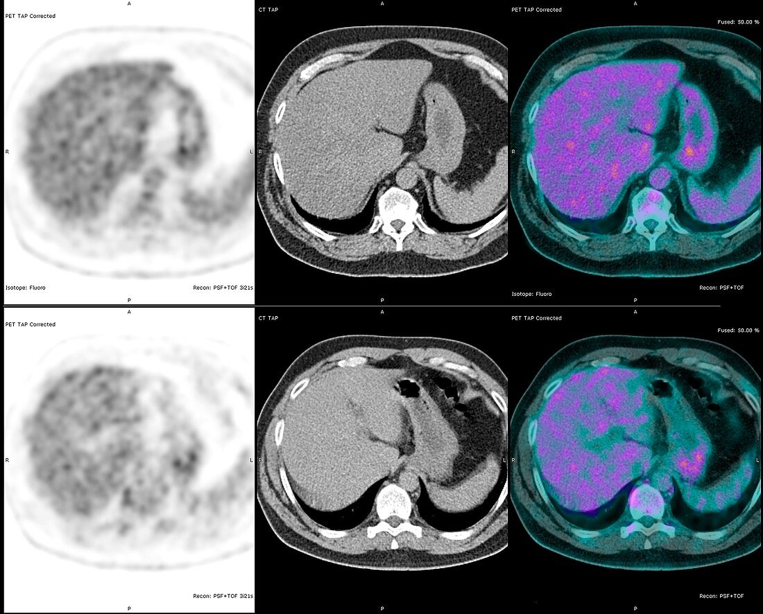
| Colon cancer scans. Axial positron emission tomography (PET) and computed tomography (CT) scans through the abdomen of a 60-year-old man with colon cancer. The PET scan is at left, the CT scan is at centre, and a combined CT-PET scan is at right. These scans (at the level of the liver) were performed to see whether the cancer has spread to other organs. The scans show no signs of secondary cancers, but do show signs of a fatty liver (hepatic steatosis). | |
| Lizenzart: | Lizenzpflichtig |
| Credit: | Science Photo Library / Zephyr |
| Bildgröße: | 4658 px × 3751 px |
| Modell-Rechte: | nicht erforderlich |
| Eigentums-Rechte: | nicht erforderlich |
| Restrictions: | - |
Preise für dieses Bild ab 15 €
Universitäten & Organisationen
(Informationsmaterial Digital, Informationsmaterial Print, Lehrmaterial Digital etc.)
ab 15 €
Redaktionell
(Bücher, Bücher: Sach- und Fachliteratur, Digitale Medien (redaktionell) etc.)
ab 30 €
Werbung
(Anzeigen, Aussenwerbung, Digitale Medien, Fernsehwerbung, Karten, Werbemittel, Zeitschriften etc.)
ab 55 €
Handelsprodukte
(bedruckte Textilie, Kalender, Postkarte, Grußkarte, Verpackung etc.)
ab 75 €
Pauschalpreise
Rechtepakete für die unbeschränkte Bildnutzung in Print oder Online
ab 495 €
Keywords
- 3,
- 60er Jahre,
- Abdomen,
- abnormal,
- axial,
- Bauch,
- Computertomographie,
- ct,
- CT-Scan,
- Darmkrebs,
- Diagnose,
- Drei,
- Einfarbig,
- Erwachsene,
- farbig,
- Fettleber,
- geduldig,
- gefärbt,
- Haustier,
- hepatisch,
- Hepatologie,
- kombiniert,
- Kondition,
- Krankheit,
- Krebs,
- krebsartig,
- Leber,
- maligne,
- Malignom,
- Mann,
- Männlich,
- Medizin,
- medizinisch,
- menschlicher Körper,
- Niemand,
- Onkologie,
- PET-CT,
- Scanner,
- Schwarz und weiß,
- schwarzer Hintergrund,
- sechziger Jahre,
- Sektion,
- sektioniert,
- Störung,
- Thorax,
- Trio,
- Tumor,
- ungesund,
- weißer Hintergrund