Brain arteries after stroke treatment, angiogram
Bildnummer 12503890
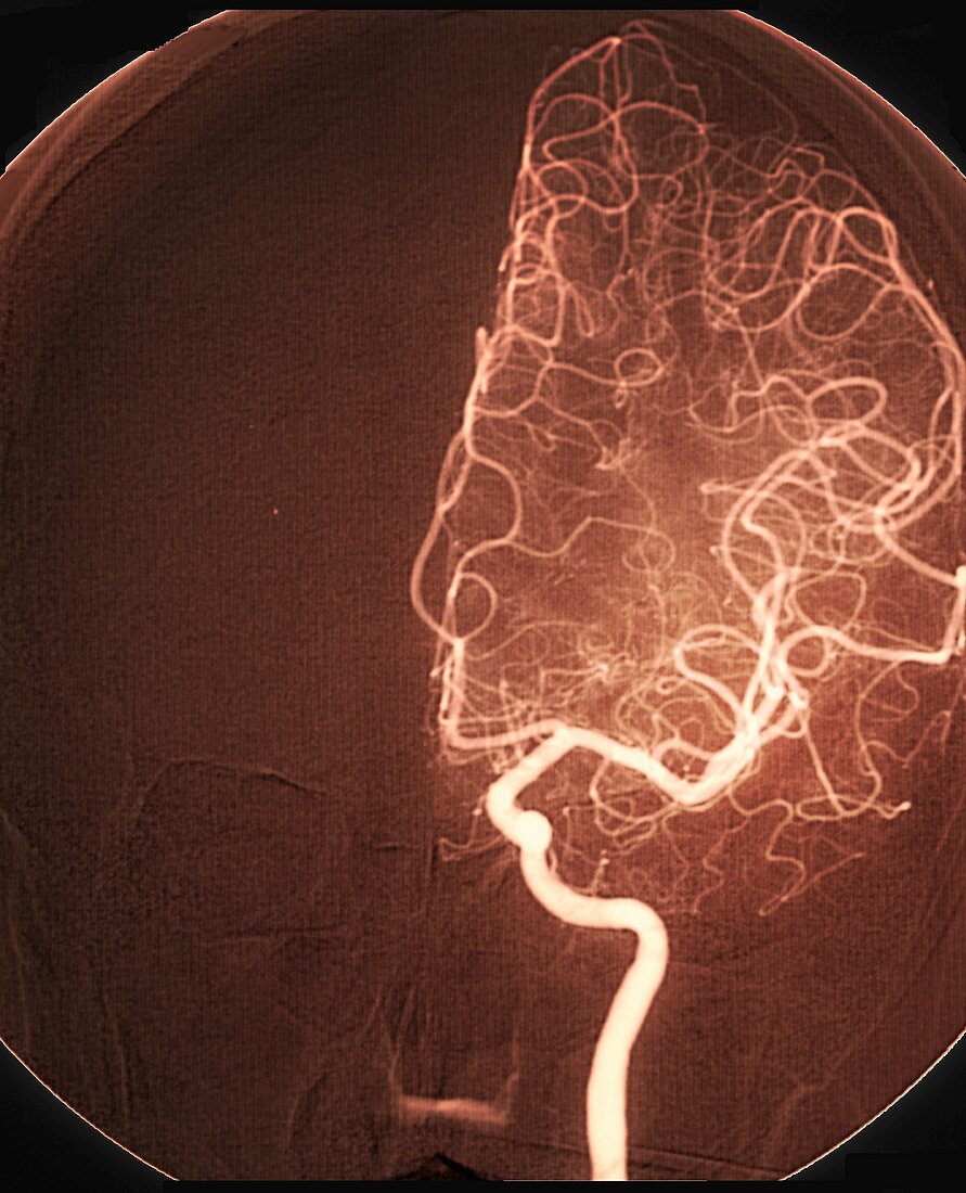
| Brain arteries after stroke treatment. Coloured angiogram of arteries in the affected area of the brain of a 54-year-old woman after treatment for a stroke (cerebrovascular accident, CVA). The stroke occurred due to a blocked (occluded) left middle cerebral artery (shown here) causing areas of ischaemia (lack of blood flow) to parts of the brain. After treatment with thrombolytic (clot-busting) drugs, blood flow was restored to the affected area. Angiograms use a contrast dye to highlight blood vessels. For X-rays showing the blocked blood vessels before treatment, see images C038/8719, C038/8729 and C038/8730 (includes arrows showing the location of the blockages). | |
| Lizenzart: | Lizenzpflichtig |
| Credit: | Science Photo Library / Zephyr |
| Bildgröße: | 3763 px × 4644 px |
| Modell-Rechte: | nicht erforderlich |
| Eigentums-Rechte: | nicht erforderlich |
| Restrictions: | - |
Preise für dieses Bild ab 15 €
Universitäten & Organisationen
(Informationsmaterial Digital, Informationsmaterial Print, Lehrmaterial Digital etc.)
ab 15 €
Redaktionell
(Bücher, Bücher: Sach- und Fachliteratur, Digitale Medien (redaktionell) etc.)
ab 30 €
Werbung
(Anzeigen, Aussenwerbung, Digitale Medien, Fernsehwerbung, Karten, Werbemittel, Zeitschriften etc.)
ab 55 €
Handelsprodukte
(bedruckte Textilie, Kalender, Postkarte, Grußkarte, Verpackung etc.)
ab 75 €
Pauschalpreise
Rechtepakete für die unbeschränkte Bildnutzung in Print oder Online
ab 495 €
Keywords
- 50er Jahre,
- abnormal,
- Angiografie,
- Angiogramm,
- Arterie,
- arteriell,
- Behandlung,
- Blutfluss,
- Blutgefäß,
- CVA,
- Diagnose,
- Drogen,
- Erwachsene,
- farbig,
- Fünfziger Jahre,
- geduldig,
- gefärbt,
- Gehirn,
- Ischämie,
- Kondition,
- Medizin,
- medizinisch,
- Mensch,
- menschlicher Körper,
- Neurologie,
- neurologisch,
- Niemand,
- Radiographie,
- Röntgen,
- Röntgengerät,
- Schlaganfall,
- Störung,
- Thrombolyse,
- ungesund,
- vaskulär