Atherosclerosis in neck arteries, 3D CT scan
Bildnummer 12493605
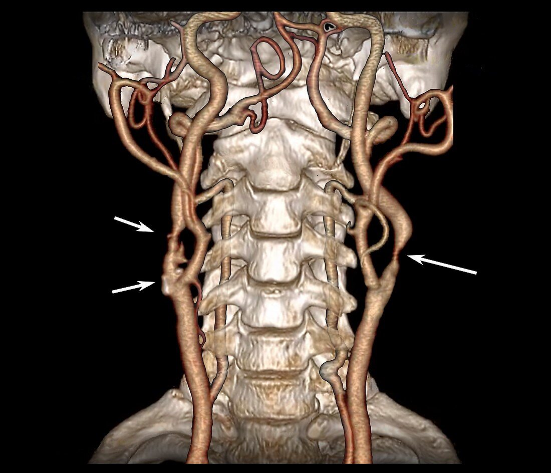
| Atherosclerosis in neck arteries. 3D coloured computed tomography (CT) scan and angiogram of the neck arteries (orange) and bones in a 59-year-old man with atherosclerosis. The scan shows the carotid blood vessels in anterior (frontal) view, with the bones of the face removed. The irregular appearance of the arterial walls, and areas of narrowing (three areas marked with arrows), indicate the presence of atheromatous plaques, deposits of cholesterol and fats on the blood vessel walls. This narrowing (stenosis) is a risk factor for strokes. | |
| Lizenzart: | Lizenzpflichtig |
| Credit: | Science Photo Library / Zephyr |
| Bildgröße: | 4513 px × 3872 px |
| Modell-Rechte: | nicht erforderlich |
| Eigentums-Rechte: | nicht erforderlich |
| Restrictions: | - |
Preise für dieses Bild ab 15 €
Universitäten & Organisationen
(Informationsmaterial Digital, Informationsmaterial Print, Lehrmaterial Digital etc.)
ab 15 €
Redaktionell
(Bücher, Bücher: Sach- und Fachliteratur, Digitale Medien (redaktionell) etc.)
ab 30 €
Werbung
(Anzeigen, Aussenwerbung, Digitale Medien, Fernsehwerbung, Karten, Werbemittel, Zeitschriften etc.)
ab 55 €
Handelsprodukte
(bedruckte Textilie, Kalender, Postkarte, Grußkarte, Verpackung etc.)
ab 75 €
Pauschalpreise
Rechtepakete für die unbeschränkte Bildnutzung in Print oder Online
ab 495 €
Keywords
- 3-dimensional,
- 3D,
- 50er Jahre,
- abnormal,
- Angiografie,
- Angiogramm,
- anterior,
- Arterie,
- arteriell,
- Arterien,
- Atherom,
- Atherosklerose,
- beschriftet,
- Blutgefäß,
- Blutgefäße,
- Computertomographie,
- CT-Scan,
- Dreidimensional,
- Erwachsene,
- Etikette,
- Etiketten,
- extern,
- farbig,
- Frontansicht,
- Fünfziger Jahre,
- geduldig,
- gefärbt,
- Hals,
- intern,
- Kondition,
- Kontrastmittel,
- Krankheit,
- Kreislauf,
- Mann,
- Männlich,
- Medizin,
- medizinisch,
- menschlicher Körper,
- Niemand,
- Scanner,
- schwarzer Hintergrund,
- Stenose,
- Stenosen,
- Störung,
- ungesund,
- vaskulär,
- verbreitet