Bamboo cane cross section, light micrograph
Bildnummer 12491823
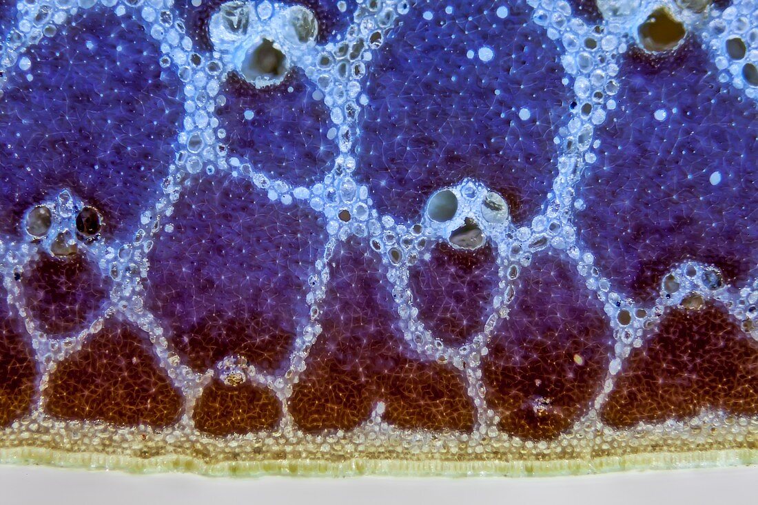
| Light micrograph of a section through a bamboo cane (diameter 2cm) used for gardening. Commercially available Bamboo canes are too hard to cut in thin layers for microscopic examination, so a small part of a bamboo cane was embedded in light curing resin and then ground and diamond polished. Several vascular bundles are visible, the dark brown cells have thick, lignified cell walls which are responsible for the stability of the bamboo canes. Image taken from the outer area of the cane with the epidermis visible at the bottom and a high density of lignified cells (fluorescing blue). Microscopic contrast technique: Incident brightfield combined with UV excitation fluorescence. Magnification: 200x when printed 10 centimetres wide. | |
| Lizenzart: | Lizenzpflichtig |
| Credit: | Science Photo Library / Guenther, Gerd |
| Bildgröße: | 5616 px × 3744 px |
| Modell-Rechte: | nicht erforderlich |
| Eigentums-Rechte: | nicht erforderlich |
| Restrictions: | - |
Preise für dieses Bild ab 15 €
Universitäten & Organisationen
(Informationsmaterial Digital, Informationsmaterial Print, Lehrmaterial Digital etc.)
ab 15 €
Redaktionell
(Bücher, Bücher: Sach- und Fachliteratur, Digitale Medien (redaktionell) etc.)
ab 30 €
Werbung
(Anzeigen, Aussenwerbung, Digitale Medien, Fernsehwerbung, Karten, Werbemittel, Zeitschriften etc.)
ab 55 €
Handelsprodukte
(bedruckte Textilie, Kalender, Postkarte, Grußkarte, Verpackung etc.)
ab 75 €
Pauschalpreise
Rechtepakete für die unbeschränkte Bildnutzung in Print oder Online
ab 495 €
Keywords
- Anatomie,
- anatomisch,
- Bambus,
- Biologie,
- biologisch,
- Botanik,
- botanisch,
- Bündel,
- Bündeln,
- bunt,
- farbig,
- Flora,
- gefärbt,
- Gefäß,
- Gefäße,
- Gefäßsystem,
- Histologie,
- histologisch,
- Lichtmikroskop,
- lichtmikroskopische Aufnahme,
- Mahlen,
- Mikroskop,
- Natur,
- Oberfläche,
- Pflanze,
- Pflanzen,
- Phloem,
- Polieren,
- Stengel,
- Struktur,
- Strukturen,
- Transport,
- Xylem