Colon cancer cells, fluorescence micrograph
Bildnummer 12450655
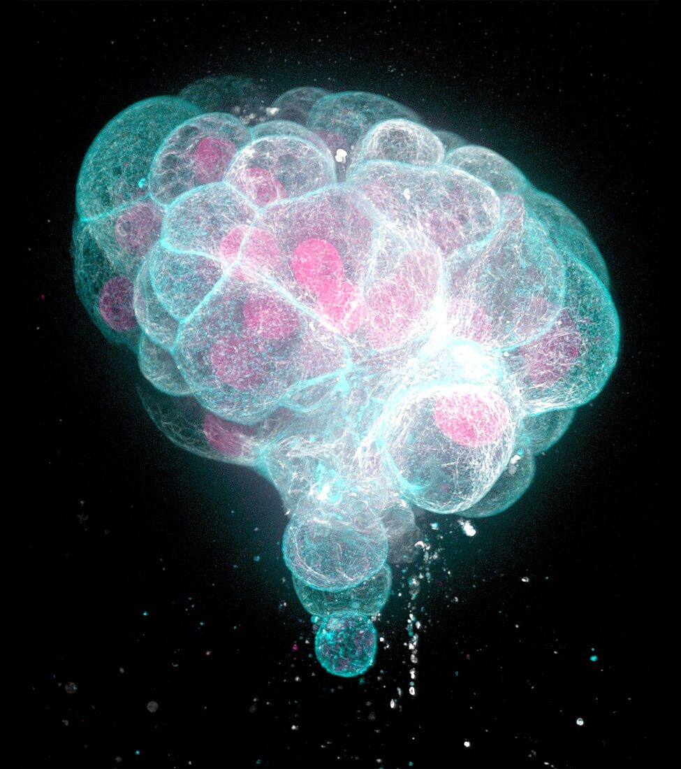
| Fluorescence micrograph of human colon cancer cells in a three-dimensional extracellular matrix. This environment mimics physiological tissue, and the cells organize into cancer organoids. Such 3D organoid cultures are used by researchers to understand the molecules and mechanisms involved in tissue formation, and in the case of cancer to understand pathological cell behavior and to develop new therapies. These cells were stained by immunofluorescence for two components of the cell cytoskeleton: Filamentous actin (cyan) that highlights microscopic cell surface structures and cell-cell contacts, and microtubules (white) that form a transport system inside cells. In addition, the DNA (deoxyribonucleic acid) in cell nuclei appears red. | |
| Lizenzart: | Lizenzpflichtig |
| Credit: | Science Photo Library / Wittmann, Dr. Torsten |
| Bildgröße: | 3047 px × 3439 px |
| Modell-Rechte: | nicht erforderlich |
| Eigentums-Rechte: | nicht erforderlich |
| Restrictions: | - |
Preise für dieses Bild ab 15 €
Universitäten & Organisationen
(Informationsmaterial Digital, Informationsmaterial Print, Lehrmaterial Digital etc.)
ab 15 €
Redaktionell
(Bücher, Bücher: Sach- und Fachliteratur, Digitale Medien (redaktionell) etc.)
ab 30 €
Werbung
(Anzeigen, Aussenwerbung, Digitale Medien, Fernsehwerbung, Karten, Werbemittel, Zeitschriften etc.)
ab 55 €
Handelsprodukte
(bedruckte Textilie, Kalender, Postkarte, Grußkarte, Verpackung etc.)
ab 75 €
Pauschalpreise
Rechtepakete für die unbeschränkte Bildnutzung in Print oder Online
ab 495 €
Keywords
- 3D,
- abnormal,
- Aktin,
- Anatomie,
- anatomisch,
- Atomkern,
- Biologie,
- biologisch,
- Desoxiribonukleinsäure,
- DNA,
- Eiweiß,
- Experiment,
- Fibroblasten,
- Fluoreszenz,
- fluoreszierend,
- Forschung,
- Gewebe,
- Gewebekultur,
- Histologie,
- histologisch,
- Kerne,
- Kondition,
- Krankheit,
- Krebs,
- krebsartig,
- Krebszelle,
- Lichtmikroskop,
- lichtmikroskopische Aufnahme,
- maligne,
- Malignom,
- Medizin,
- medizinisch,
- Metastase,
- Mikrotubuli,
- Modell-,
- Niemand,
- Onkologie,
- schwarzer Hintergrund,
- Störung,
- Tumor,
- ungesund,
- Zellbilogie,
- Zelle,
- Zellen,
- zellular,
- Zytoskelett