Pelvis and lumbar spine, 3D CT scan
Bildnummer 12450651
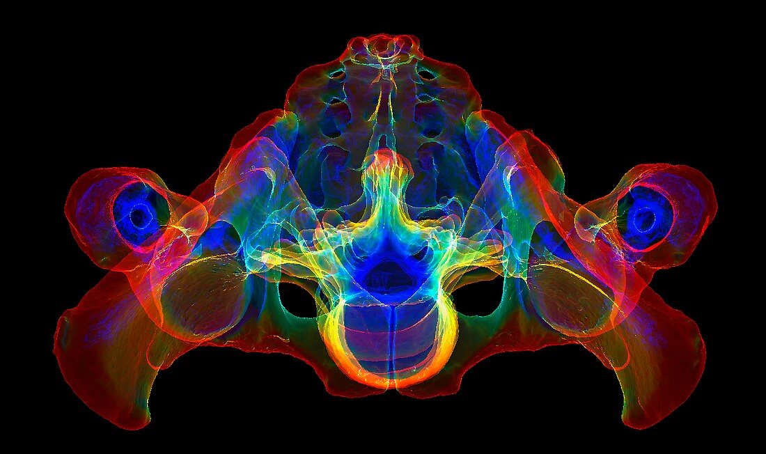
| Pelvis and lumbar spine. Coloured 3D computed tomography (CT) scan from below (inferior view) of the normal human pelvis and lumbar spine. The spinal vertebrae are at lower centre. Sectioned views of the femurs (upper leg bones) are seen at left and right, articulating with the bones of the pelvis to form the hip joints. | |
| Lizenzart: | Lizenzpflichtig |
| Credit: | Science Photo Library / Fung, K.H. |
| Bildgröße: | 6000 px × 3557 px |
| Modell-Rechte: | nicht erforderlich |
| Eigentums-Rechte: | nicht erforderlich |
| Restrictions: | - |
Preise für dieses Bild ab 15 €
Universitäten & Organisationen
(Informationsmaterial Digital, Informationsmaterial Print, Lehrmaterial Digital etc.)
ab 15 €
Redaktionell
(Bücher, Bücher: Sach- und Fachliteratur, Digitale Medien (redaktionell) etc.)
ab 30 €
Werbung
(Anzeigen, Aussenwerbung, Digitale Medien, Fernsehwerbung, Karten, Werbemittel, Zeitschriften etc.)
ab 55 €
Handelsprodukte
(bedruckte Textilie, Kalender, Postkarte, Grußkarte, Verpackung etc.)
ab 75 €
Pauschalpreise
Rechtepakete für die unbeschränkte Bildnutzung in Print oder Online
ab 495 €
Keywords
- Anatomie,
- anatomisch,
- Arthrologie,
- ausgeschnitten,
- Ausschnitte,
- Becken,
- Beine,
- Biologie,
- biologisch,
- Computertomographie,
- CT-Scan,
- farbig,
- Femur,
- gefärbt,
- Gelenk,
- Gelenke,
- gesund,
- Hüfte,
- Hüften,
- Joint,
- Knochen,
- Kreuzbein,
- Lendenwirbelsäule,
- menschlicher Körper,
- Niemand,
- normal,
- Osteologie,
- Sakral,
- Scanner,
- schwarzer Hintergrund,
- Steißbein,
- Wirbel,
- Wirbelsäule,
- Wirbelsäulen-