Brain blood vessel, fluorescence light micrograph
Bildnummer 12450605
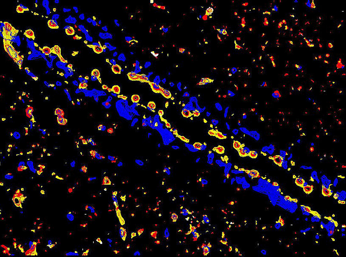
| Brain blood vessel. Fluorescence deconvolution light micrograph of a blood vessel from the brain of an animal being used to model neurodegeneration. The red is TNF-alpha (tumor necrosis factor-alpha). The yellow is co-localization of TNF and the regulatory protein ubiquitin. Cell nuclei (blue) have been highlighted with a DAPI stain. | |
| Lizenzart: | Lizenzpflichtig |
| Credit: | Science Photo Library / R. BICK, B. POINDEXTER, UT MEDICAL SCHOOL |
| Bildgröße: | 3428 px × 2549 px |
| Modell-Rechte: | nicht erforderlich |
| Eigentums-Rechte: | nicht erforderlich |
| Restrictions: | - |
Preise für dieses Bild ab 15 €
Universitäten & Organisationen
(Informationsmaterial Digital, Informationsmaterial Print, Lehrmaterial Digital etc.)
ab 15 €
Redaktionell
(Bücher, Bücher: Sach- und Fachliteratur, Digitale Medien (redaktionell) etc.)
ab 30 €
Werbung
(Anzeigen, Aussenwerbung, Digitale Medien, Fernsehwerbung, Karten, Werbemittel, Zeitschriften etc.)
ab 55 €
Handelsprodukte
(bedruckte Textilie, Kalender, Postkarte, Grußkarte, Verpackung etc.)
ab 75 €
Pauschalpreise
Rechtepakete für die unbeschränkte Bildnutzung in Print oder Online
ab 495 €
Keywords
- abnormal,
- Aktin,
- Anatomie,
- anatomisch,
- Atomkern,
- Biologie,
- biologisch,
- Blutgefäß,
- dapi,
- Degeneration,
- Eiweiß,
- Fluoreszenzentfaltung,
- Fluoreszenzlichtmikroskopische Aufnahme,
- fluoreszierend,
- Gehirn,
- Gewebe,
- Histologie,
- histologisch,
- Kerne,
- Kondition,
- Krankheit,
- Kreislauf,
- Lichtmikroskop,
- lichtmikroskopische Aufnahme,
- Medizin,
- medizinisch,
- menschlicher Körper,
- Mikroskopie,
- Neurologie,
- neurologisch,
- Niemand,
- Proteine,
- schwarzer Hintergrund,
- Störung,
- Tierkörper,
- tnf,
- ungesund,
- vaskulär,
- Zellbilogie,
- Zelle,
- Zellen,
- Zytologie,
- Zytologisch