Intestinal lining, SEM
Bildnummer 12395208
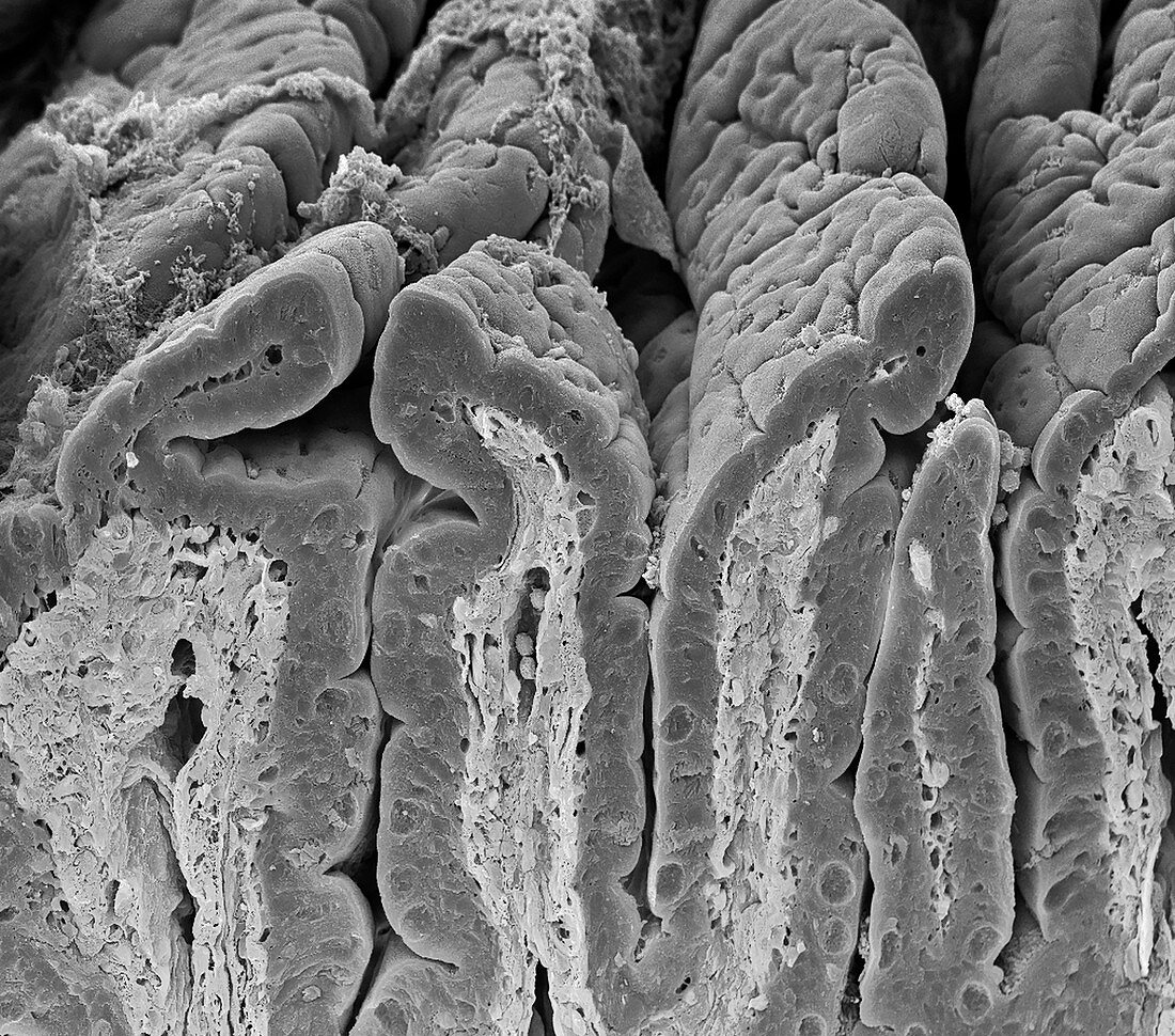
| Intestinal lining. Scanning electron micrograph (SEM) of a freeze-fractured surface of the small intestine. The surface consists of deep folds, called villi, that have been sectioned in this view. The intestinal surface (orange, upper frame) is exposed to food. The underlying structure of this surface is seen in the sectioned area. Surface (epithelial) cells are supported by connective tissue that forms the core of each fold (villus). The folds increase the area for the absorption of nutrients from food. The height of a villus varies in a small intestine from 0.3 to 0.8 millimetres. Magnification: x 250 at 10 centimetres wide. | |
| Lizenzart: | Lizenzpflichtig |
| Credit: | Science Photo Library / Gschmeissner, Steve |
| Bildgröße: | 4572 px × 4031 px |
| Modell-Rechte: | nicht erforderlich |
| Eigentums-Rechte: | nicht erforderlich |
| Restrictions: | - |
Preise für dieses Bild ab 15 €
Universitäten & Organisationen
(Informationsmaterial Digital, Informationsmaterial Print, Lehrmaterial Digital etc.)
ab 15 €
Redaktionell
(Bücher, Bücher: Sach- und Fachliteratur, Digitale Medien (redaktionell) etc.)
ab 30 €
Werbung
(Anzeigen, Aussenwerbung, Digitale Medien, Fernsehwerbung, Karten, Werbemittel, Zeitschriften etc.)
ab 55 €
Handelsprodukte
(bedruckte Textilie, Kalender, Postkarte, Grußkarte, Verpackung etc.)
ab 75 €
Pauschalpreise
Rechtepakete für die unbeschränkte Bildnutzung in Print oder Online
ab 495 €
Keywords
- Anatomie,
- anatomisch,
- Biologie,
- biologisch,
- Darm,
- Dünndarm,
- Einfarbig,
- Einfrieren,
- Epithel,
- epithelial,
- Falten,
- frakturiert,
- Futter,
- Gefaltet,
- gefriergebrochen,
- gesund,
- Gewebe,
- Histologie,
- histologisch,
- Kelch,
- Lebensmittel,
- Mauer,
- menschlicher Körper,
- normal,
- rasterelektronenmikroskopische Aufnahme,
- REM,
- schwarz und weiß,
- sekretorisch,
- Sektion,
- sektioniert,
- Trümmer,
- Verdauung,
- Verdauungssystem,
- Zelle,
- Zellen,
- Zotte,
- Zotten