Human hip bones, illustration
Bildnummer 12394208
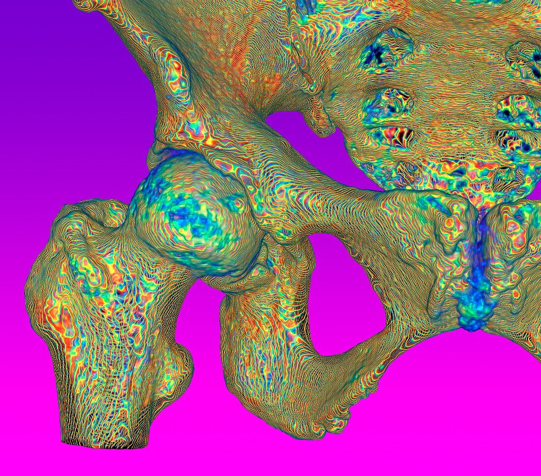
| Human hip bones. Illustration based on a 3D computed tomography (CT) scan of the bones of a right-hand human hip, seen from the front. At lower left is the femur (thigh bone). The head of the femur (ball-shaped) articulates with the acetabulum, the hip socket in the pelvis. The pelvis bones that form part of the hip joint are the ischium and pubis (centre and lower right) and the ilium (upper left). | |
| Lizenzart: | Lizenzpflichtig |
| Credit: | Science Photo Library / Fung, K.H. |
| Bildgröße: | 4487 px × 3953 px |
| Modell-Rechte: | nicht erforderlich |
| Eigentums-Rechte: | nicht erforderlich |
| Restrictions: | - |
Preise für dieses Bild ab 15 €
Universitäten & Organisationen
(Informationsmaterial Digital, Informationsmaterial Print, Lehrmaterial Digital etc.)
ab 15 €
Redaktionell
(Bücher, Bücher: Sach- und Fachliteratur, Digitale Medien (redaktionell) etc.)
ab 30 €
Werbung
(Anzeigen, Aussenwerbung, Digitale Medien, Fernsehwerbung, Karten, Werbemittel, Zeitschriften etc.)
ab 55 €
Handelsprodukte
(bedruckte Textilie, Kalender, Postkarte, Grußkarte, Verpackung etc.)
ab 75 €
Pauschalpreise
Rechtepakete für die unbeschränkte Bildnutzung in Print oder Online
ab 495 €
Keywords
- 3D,
- Anatomie,
- anatomisch,
- anterior,
- Arthrologie,
- Becken,
- Bein,
- Biologie,
- biologisch,
- Computertomographie,
- CT-Scan,
- Dreidimensional,
- farbig,
- Femur,
- Frontansicht,
- gefärbt,
- Gelenk,
- gesund,
- Hüfte,
- Joint,
- Knochen,
- Kreuzbein,
- Lila Hintergrund,
- menschlicher Körper,
- Niemand,
- normal,
- Osteologie,
- pelvin,
- Scanner,
- Skelett-,
- Steißbein