Two-horned uterus, axial pelvic CT scan
Bildnummer 12378419
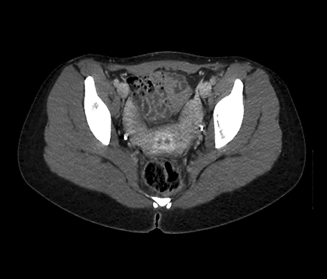
| Two-horned uterus. Axial pelvic computed tomography (CT) scan showing the pelvic organs of a 27-year-old woman with a bicornuate (two-horned) uterus. This congenital defect results in the division of the upper end of the uterus into two halves. It can affect the success of a pregnancy. Surgical treatments include reshaping the uterus, or stitching shut the neck of the uterus during pregnancy. Here, there is also an associated vaginal duplicity, indicating a possible uterus didelphys (double uterus). | |
| Lizenzart: | Lizenzpflichtig |
| Credit: | Science Photo Library / Zephyr |
| Bildgröße: | 4520 px × 3866 px |
| Modell-Rechte: | nicht erforderlich |
| Eigentums-Rechte: | nicht erforderlich |
| Restrictions: | - |
Preise für dieses Bild ab 15 €
Universitäten & Organisationen
(Informationsmaterial Digital, Informationsmaterial Print, Lehrmaterial Digital etc.)
ab 15 €
Redaktionell
(Bücher, Bücher: Sach- und Fachliteratur, Digitale Medien (redaktionell) etc.)
ab 30 €
Werbung
(Anzeigen, Aussenwerbung, Digitale Medien, Fernsehwerbung, Karten, Werbemittel, Zeitschriften etc.)
ab 55 €
Handelsprodukte
(bedruckte Textilie, Kalender, Postkarte, Grußkarte, Verpackung etc.)
ab 75 €
Pauschalpreise
Rechtepakete für die unbeschränkte Bildnutzung in Print oder Online
ab 495 €
Keywords
- 20er Jahre,
- Abdomen,
- abnormal,
- ausgeschnitten,
- Ausschnitte,
- axial,
- Bauch,
- Computertomographie,
- CT-Scan,
- Deformität,
- Diagnose,
- Einfarbig,
- Erwachsene,
- Frau,
- geduldig,
- Kondition,
- Medizin,
- medizinisch,
- menschlicher Körper,
- Niemand,
- Scanner,
- Schwarz und weiß,
- schwarzer Hintergrund,
- Störung,
- ungesund,
- Uterus,
- Vagina,
- Weiblich,
- zwanziger Jahre