Wall of a large vein, light micrograph
Bildnummer 12360993
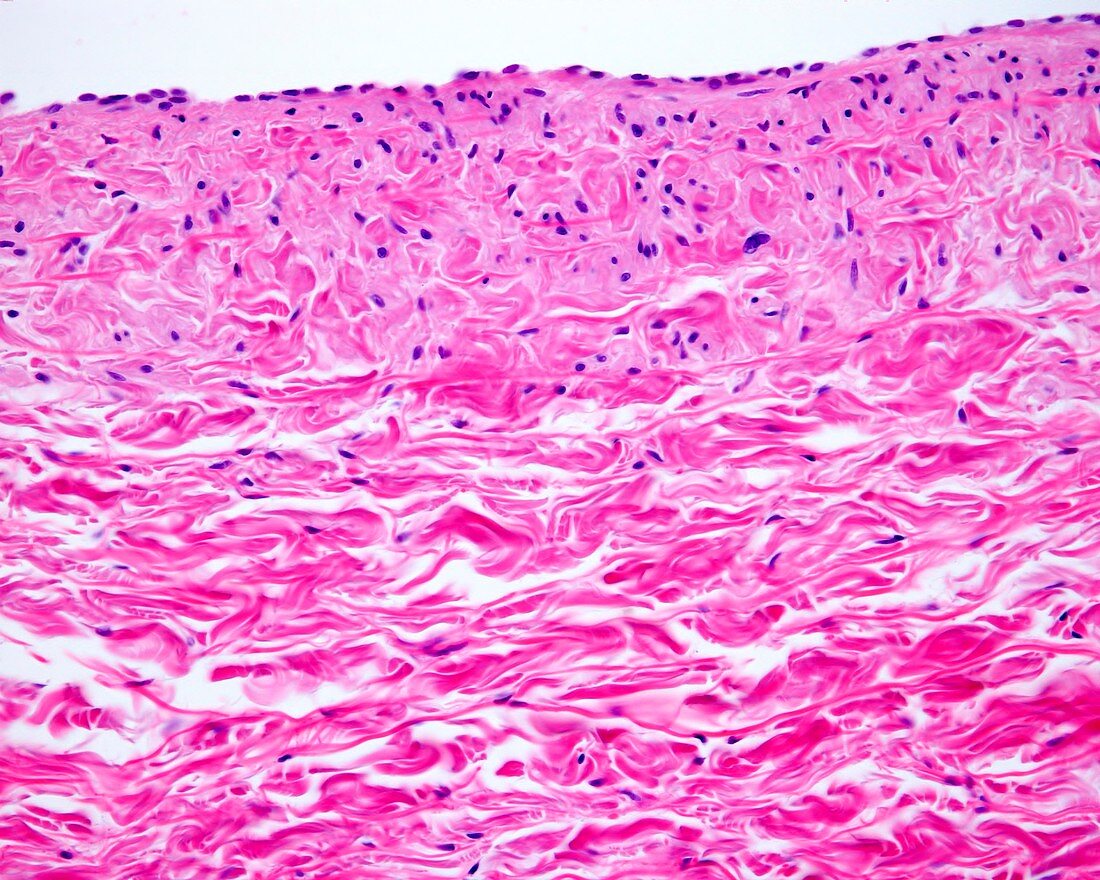
| Light micrograph of a longitudinal section of a large vein showing the layers of its wall. From the lumen, located at the top of the image, we observe: a tunica intima with the endothelium and a thin subendothelial connective tissue. A medium layer formed by a few bundles of circular cross sectioned myocytes. The majority of the wall is occupied by intermingled bundles of collagen fibres belonging to the tunica adventitia. Magnification: x180 when printed at 10 centimetres across. | |
| Lizenzart: | Lizenzpflichtig |
| Credit: | Science Photo Library / JOSE CALVO |
| Bildgröße: | 4674 px × 3739 px |
| Modell-Rechte: | nicht erforderlich |
| Eigentums-Rechte: | nicht erforderlich |
| Restrictions: | - |
Preise für dieses Bild ab 15 €
Universitäten & Organisationen
(Informationsmaterial Digital, Informationsmaterial Print, Lehrmaterial Digital etc.)
ab 15 €
Redaktionell
(Bücher, Bücher: Sach- und Fachliteratur, Digitale Medien (redaktionell) etc.)
ab 30 €
Werbung
(Anzeigen, Aussenwerbung, Digitale Medien, Fernsehwerbung, Karten, Werbemittel, Zeitschriften etc.)
ab 55 €
Handelsprodukte
(bedruckte Textilie, Kalender, Postkarte, Grußkarte, Verpackung etc.)
ab 75 €
Pauschalpreise
Rechtepakete für die unbeschränkte Bildnutzung in Print oder Online
ab 495 €
Keywords
- Adventitia,
- Anatomie,
- anatomisch,
- befleckt,
- Biologie,
- biologisch,
- Blut,
- Blutgefäß,
- Gefäß,
- Gefäße,
- Gefäßsystem,
- gesund,
- Gewebe,
- Glatt,
- Histologie,
- histologisch,
- Kreislauf,
- lichtmikroskopische Aufnahme,
- Lumen,
- Mauer,
- Medien,
- menschlicher Körper,
- Mikrofotografie,
- Mikroskopie,
- mikroskopisch,
- Muskulös,
- Schicht,
- Sektion,
- sektioniert,
- vaskulär,
- Vene,
- venös,
- Verfärbung